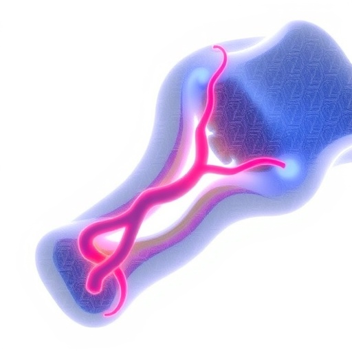
Penn State researchers have developed a 3D bioprinting strategy that uses genetically guided clusters of living stem cells to promote the regeneration of damaged bone while encouraging the growth of new blood vessels. In laboratory experiments and studies involving immunodeficient mice, the team showed that carefully engineered cellular spheroids could be positioned inside a supportive microgel scaffold and directed toward different biological functions. Some spheroids were optimized for bone formation, while others were programmed to support vascularization, the process through which new blood vessels develop inside regenerating tissue. The findings establish a potential framework for treating severe bone loss caused by major trauma, cancer or infection—conditions in which conventional healing may be insufficient.
The work addresses one of the central challenges in bone tissue engineering. Bone is not simply a rigid mineralized structure; it is a living, highly organized tissue supplied by an intricate network of blood vessels. When an engineered bone graft becomes too thick, cells located far from existing circulation can lack oxygen and nutrients, limiting their survival and preventing the tissue from maturing. Without a functional vascular network, even a scaffold containing appropriate cells and growth signals may fail to integrate with the body. The Penn State team therefore sought to create a cellular system in which bone development and blood-vessel formation could occur together rather than as separate stages.
The researchers began with commercially available, undifferentiated stem cells. These cells have the capacity to develop into more specialized cell types, but their fate must be controlled through chemical, genetic or environmental signals. The team introduced two microRNA molecules, miR-148b and miR-210, into separate populations of cells. MicroRNAs are short strands of noncoding genetic material that regulate gene activity by influencing the stability or translation of messenger RNA. Rather than encoding proteins themselves, they can act as molecular switches that adjust networks of genes involved in cell differentiation and tissue development. In this study, miR-148b was associated with pathways supporting bone formation, while miR-210 was used to promote characteristics linked to vascular development.
After the cells were exposed to the microRNAs and cultured for several days, they organized into spheroids—compact, three-dimensional clusters of living cells. Spheroids are increasingly used in tissue engineering because they can reproduce some of the cell-to-cell interactions found in natural tissues more effectively than isolated cells suspended in a solution. Within these clusters, cells communicate through direct contact and secreted signaling molecules, creating local biochemical environments that can influence maturation. The researchers produced multiple spheroid types, including clusters treated with only one microRNA and clusters containing cells influenced by both pathways. They then combined these populations in defined arrangements to determine whether cells with complementary functions would cooperate during tissue regeneration.
To place the spheroids with the necessary precision, the team used aspiration-assisted bioprinting, a technique developed in the laboratory of biomedical engineering professor Ibrahim Ozbolat. The method uses controlled suction to collect individual spheroids and position them at predetermined locations inside a soft microgel-based scaffold. Unlike conventional bioprinting approaches that deposit a continuous stream of cell-laden bioink, aspiration-assisted printing can arrange larger living cell aggregates one by one. This allows researchers to control the distance between spheroids, their spatial pattern and the relationships among different cell populations. Uniform spacing is important because it can help establish more consistent signaling and reduce the risk that some clusters become isolated from nutrients or neighboring cells.
The scaffold served as a temporary three-dimensional environment in which the printed spheroids could survive, mature and interact. Researchers allowed the constructs to culture for 28 days before analyzing their gene activity and cellular markers. This period gave the microRNA-directed cells time to progress along differentiation pathways while remaining in a structured arrangement. The team examined markers associated with bone formation and vascular development to determine how strongly each spheroid type expressed its intended biological program. Constructs containing alternating or combined populations were particularly important because they were designed to reproduce the cooperation that occurs between different cell types during natural tissue growth.
The researchers also implanted the printed constructs into immunodeficient mice with bone defects and monitored regeneration for six weeks. The comparison revealed a substantial difference between untreated animals and animals receiving a control spheroid construct. In mice that received no treatment, newly regenerated bone covered approximately 35 percent of the damaged region after six weeks. In animals treated with the control spheroid scaffold, coverage reached about 93 percent, suggesting that the physical presence of the scaffold and organized cell clusters alone could strongly stimulate repair. Constructs containing the microRNA-directed spheroids produced additional improvements in overall bone development and vascularization, with the combination of miR-148b- and miR-210-related populations showing the most promising performance.
One notable result involved CD31, a protein commonly used as a marker of endothelial cells and the inner lining of blood vessels. The constructs containing the combined microRNA strategy showed higher CD31 expression than both nontransfected spheroids and constructs designed around only one specialized pathway. This suggests that the interaction between differently programmed cell populations may have enhanced the formation of vascular structures. The precise mechanism remains uncertain. Cells optimized for bone development may have altered the local matrix or released signals that supported vascular cells, while the vascularly directed cells may have improved oxygen delivery and created conditions favorable to mineralized tissue formation. The researchers emphasized that the relationship between blood-vessel growth and bone development is complex and requires further investigation.
The findings do not represent an immediate treatment for ordinary fractures, and the experiments remain at a preclinical stage. The approach is intended for difficult cases involving extensive bone loss, such as injuries that remove large sections of tissue or damage caused by tumors and severe infections. Before clinical use, researchers will need to determine how the printed constructs behave in larger animal models, how long the newly formed vessels remain functional and whether vascularization could sometimes interfere with the maturation or organization of bone. They will also need to assess manufacturing consistency, immune responses and the long-term safety of introducing microRNA-modified cells. Nevertheless, the study demonstrates how precise cellular positioning and temporary genetic instructions can be combined to build more biologically coordinated grafts. By printing spheroids with distinct but complementary functions, the Penn State team is moving bioprinting closer to the goal of producing living tissues that can integrate with the body rather than merely fill a defect.
Subject of Research: Animals
Article Title: Bioprinting of miRNA-induced spheroids for vascularized, heterocellular bone regeneration
News Publication Date: 19-Jun-2026
Web References: https://doi.org/10.1016/j.cej.2026.178521
References: Chemical Engineering Journal, DOI: 10.1016/j.cej.2026.178521
Image Credits: Provided by Ibrahim Ozbolat / Daniel Hayes
Keywords
3D bioprinting, bone regeneration, tissue engineering, vascularization, spheroids, microRNA, miR-148b, miR-210, stem cells, aspiration-assisted bioprinting, regenerative medicine, biomedical engineering, tissue scaffolds, bone tissue engineering, cell transplantation
Tags: 3D bioprinting of vascularized bone tissueadvanced bone tissue repair strategiesbiofabrication of living bone graftsengineered tissue constructsgenetically guided stem cell clusters for bone regenerationgenetically programmed tissue scaffoldsin vivo studies of bioprinted bone with blood vesselsmicrogel scaffolds for tissue engineeringovercoming nutrient diffusion limitations in tissue engineeringprogramming cellular spheroids for vascularizationregenerative medicine for severe bone lossstem cell-based blood vessel growth in bioprinted tissues