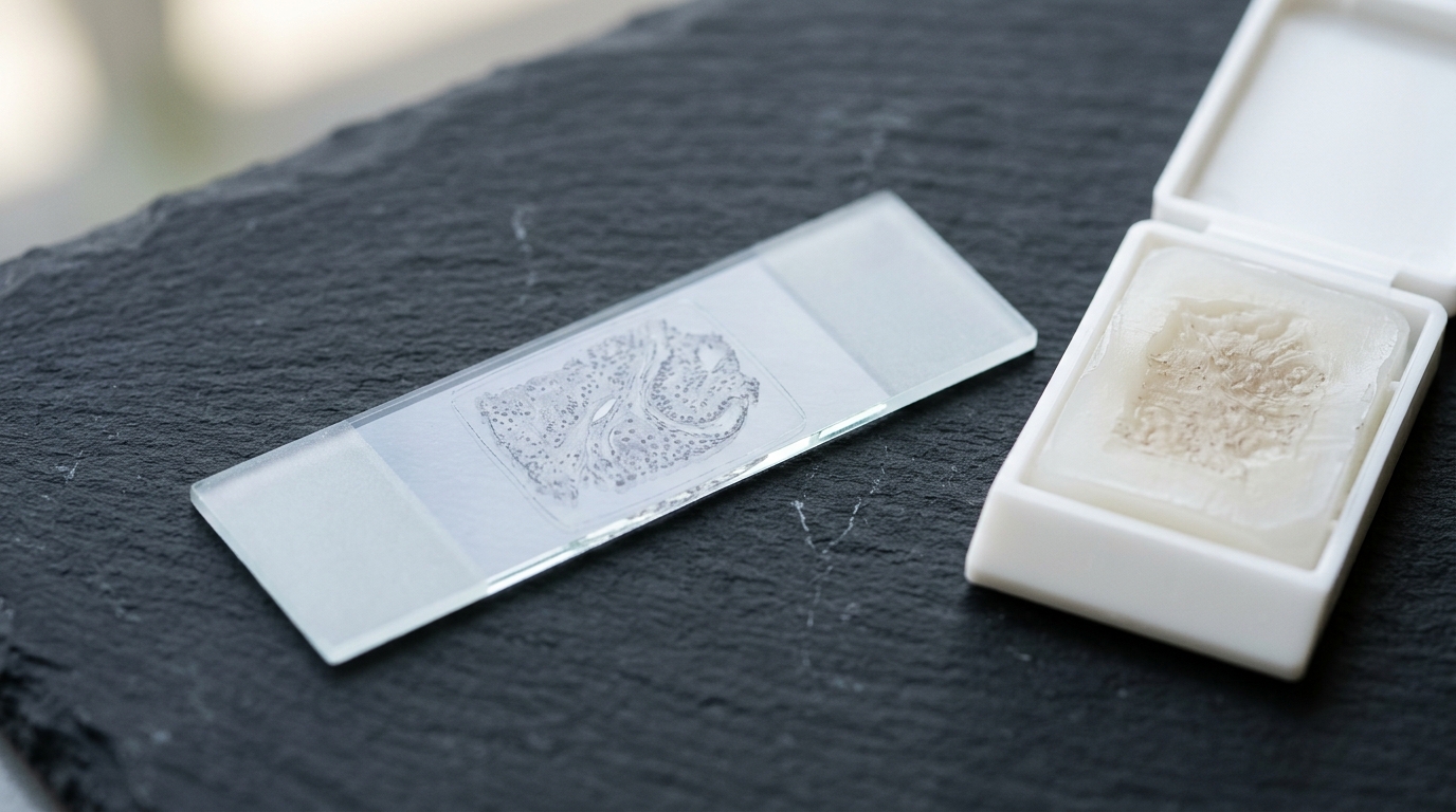
A newly published methodology establishes an adaptable and highly efficient sample preparation workflow that enables deep spatial proteome profiling from minimal amounts of archival tissue without requiring specialized proprietary hardware. Developed for laser capture microdissection on the Zeiss PALM Microbeam instrument, the streamlined protocol extracts extensive molecular information from low-input formalin-fixed, paraffin-embedded specimens, matching or exceeding the proteomic depth achieved by earlier platforms. Formalin-fixed archiving represents the gold standard for preserving clinical specimens across hospital pathology departments worldwide, maintaining tissue architecture for retrospective research. However, extensive chemical crosslinking introduced by formalin fixation traditionally creates severe barriers to extracting intact proteins, hindering molecular investigations. Previous attempts to overcome these extraction hurdles on the Zeiss platform depended heavily on specialized protocols and customized hardware components, such as nanoPOTS, which restricted accessibility and broad standardization across diagnostic laboratories. The new workflow resolves these limitations by introducing a simplified collection and processing sequence that utilizes standard off-the-shelf laboratory consumables.
To resolve tissue handling and extraction bottlenecks, the researchers systematically evaluated multiple lysis and collection buffers alongside mechanical processing variables using dense human pancreatic exocrine acinar tissue. The investigation tested harsh chemical lysing formulations, including guanidinium chloride and hydrogen peroxide, against milder solutions such as phosphate-buffered saline and triethylammonium bicarbonate. Unexpectedly, milder solutions outperformed harsher lysis agents, and the LC-MS-compatible buffer triethylammonium bicarbonate generated the highest overall protein yields. The researchers also examined the chemical addition of tris(2-carboxyethyl)phosphine hydrochloride for disulfide reduction and chloroacetamide for alkylation. Contrary to common extraction practices, incorporating these reducing and alkylating agents into triethylammonium bicarbonate and phosphate-buffered saline reduced total protein identifications. Detailed peptide-level examinations revealed that omitting alkylation had no detrimental impact on missed enzymatic cleavages, although it produced a reduction in cysteine-containing peptides. Because triethylammonium bicarbonate is completely compatible with downstream mass spectrometry and introduced no contaminating elements, it was established as the primary extraction buffer.
Because triethylammonium bicarbonate is intrinsically compatible with liquid chromatography and tandem mass spectrometry, the authors investigated whether standard solid-phase clean-up procedures could be eliminated entirely. Evaluating the workflow with and without single-pot, solid-phase-enhanced sample preparation demonstrated that omitting the clean-up step led to substantial gains in protein recovery. Specifically, bypassing solid-phase extraction produced an approximate fifty-five percent increase in identified proteins from fifty thousand square micrometers of tissue and a seventy percent increase from one hundred thousand square micrometers. Crucially, clean-up omission caused no degradation in analytical column performance, confirming that sample losses associated with solid-phase purification could be safely avoided. To address any particulate contaminants originating from the formalin-fixed specimens, the protocol relies on the C18 material housed within an EvoTip to serve as an in-line filter. Furthermore, a disposable peptide trapping cartridge was integrated into the chromatography setup to prevent non-compatible debris from entering the primary analytical column, safeguarding chromatographic stability.
Thermal reversal of formalin crosslinks and physical sample disruption were systematically evaluated to establish minimal necessary handling times. Comparing different boiling durations demonstrated that introducing a five-minute boil at ninety-five degrees Celsius increased protein identifications by approximately twenty-five percent compared to unheated controls. Extending boiling beyond five minutes produced no substantial increase in proteome coverage, indicating that the vast majority of crosslinks were reversed within that initial five-minute window. Physical disruption via sonication on a Bioruptor Pico device maintained at four degrees Celsius demonstrated minimal impact on total protein numbers, but implementing ten cycles of thirty seconds on and thirty seconds off significantly improved reproducibility across replicates. The researchers also explored a recently proposed quality-control metric tracking the relative frequency of peptides terminating in lysines versus arginines, though this metric provided no clear evidence within their experimental dataset. Additional tests evaluating altered trypsin concentrations and droplet volumes showed no measurable improvements, confirming that five minutes of boiling with ten sonication cycles provided optimal processing efficiency.
Physical recovery of tissue fragments following laser microdissection required substantial optimization because initial experiments using commercial collection tubes with adhesive-filled caps failed due to excessive tissue adherence that prevented protein extraction. To overcome this adhesion barrier, the researchers adopted a hanging drop collection strategy inspired by microfluidic principles. Twenty microliters of one hundred millimolar triethylammonium bicarbonate dissolved in high-purity water was placed directly onto the interior lid of a zero-point-five milliliter Protein LoBind tube. This volume was determined to be the minimum quantity necessary to completely cover the lid surface, ensuring that catapulted tissue fragments reliably struck the fluid droplet. Following microbeam collection, tube lids were visually inspected with a DinoLite digital microscope to verify sample capture, and misaligned tissue fragments could be corrected by gently stirring the droplet with a pipette tip. Tubes were subsequently centrifuged at twenty-one thousand three hundred times gravity for five minutes to drive collected tissue into the tube base.
Enzymatic digestion and automated loading were engineered to minimize sample handling and manual transfer losses prior to mass spectrometry. Digestion was initiated by adding ten microliters of triethylammonium bicarbonate containing one nanogram of Trypsin and Lys-C Protease Mix per twenty-five thousand square micrometers of tissue, with a baseline of two nanograms applied to sample areas under fifty thousand square micrometers. Digestion proceeded overnight at thirty-seven degrees Celsius with rotational shaking at one thousand revolutions per minute. Following digestion, samples were loaded onto activated EvoTips containing C18 material conditioned with acetonitrile, 2-propanol, and water containing zero-point-one percent formic acid. Centrifugation at seven hundred times gravity drew samples through the tips, which were then washed with acidic aqueous buffer. To prevent bound peptides from drying during batch processing, the tips were loaded with two hundred and fifty microliters of buffer and stored in a box partially filled with liquid until automated sample introduction into the chromatography system.
Peptide separation and mass spectrometric analysis were executed using high-resolution instrumentation configured for ultra-low sample amounts. Separation was performed on an EvoSep One system using the Whisper 20 method, operating at a flow rate of one hundred nanoliters per minute over a fifty-eight-minute gradient, corresponding to twenty samples per day. Peptides were resolved on a fifteen-centimeter by seventy-five-micrometer column packed with one-point-seven-micrometer C18 beads. To mitigate occasional overpressures caused by residual formalin-fixed paraffin-embedded debris accumulating on the sample collection needle, operators regularly wiped the needle every forty to sixty injections with acetonitrile-wetted cleanroom polyester wipers. The chromatography platform was coupled to an Orbitrap Eclipse Tribrid mass spectrometer through an EasySpray ion source and a FAIMSPro differential ion mobility interface operated at negative forty-five volts. Data acquisition utilized the wide isolation window high-resolution MS1 data-independent acquisition approach. In comparative trials, substituting the Eclipse Tribrid with a Thermo Orbitrap Astral mass spectrometer increased total protein identifications by approximately sixty-seven percent.
To assess analytical sensitivity and dynamic range across varying tissue quantities, the researchers benchmarked the workflow on exocrine pancreatic acinar cells ranging from one thousand one hundred and sixty-six to eight hundred thousand square micrometers, representing estimated cell numbers between two and one thousand three hundred and ten cells by volume. The authors also compared extraction efficiencies between tissue sections stained with standard hematoxylin-eosin and sections immunostained for the epithelial cell adhesion molecule EpCAM. Across tested inputs, EpCAM-stained tissue yielded slightly higher identifications, recovering approximately one thousand two hundred unique protein groups at the lowest input and expanding to approximately five thousand nine hundred protein groups at the highest input. Samples derived from hematoxylin-eosin-stained sections identified approximately nine hundred protein groups at the minimum input and reached approximately five thousand two hundred groups at the maximum. Because both staining preparations approached an identification plateau near two hundred thousand square micrometers, an input of fifty thousand to one hundred thousand square micrometers—corresponding to approximately eighty-two cells—was identified as the optimal analytical window.
The biological utility of the optimized protocol was demonstrated across three distinct malignancies: pancreatic ductal adenocarcinoma, lung adenocarcinoma, and glioblastoma. Using fifty thousand square micrometers of tissue per sample, the workflow generated consistent protein identification totals across distinct tumor types while maintaining low protein-level coefficients of variation and acceptable missed cleavage frequencies across biological replicates. Principal component analysis and hierarchical clustering revealed distinct segregation among the three cancer types, reflecting tissue-specific protein expression patterns. Differential expression analysis highlighted prominent tumor-specific markers, including significant elevation of SELENBP1 in lung adenocarcinoma and S100P in both lung and pancreatic adenocarcinoma. Evaluating identified proteins against functional reference libraries demonstrated extensive coverage of therapeutic targets, including transcription factors, epigenetic regulators, surface proteins, and protein kinases. Furthermore, Gene Ontology enrichment analysis conducted on kinases identified within pancreatic cancer samples revealed statistically significant enrichment in crucial signaling pathways, including MAPK, ErbB, vascular endothelial growth factor, and insulin signaling networks.
Direct comparisons with existing literature highlight the quantitative efficiency of this non-proprietary spatial proteomics method on archival specimens. While direct benchmarking across platforms is complicated by varying instrument setups, the closest published methodology on the Zeiss PALM Microbeam platform by Davis and colleagues in 2023 identified nearly three thousand proteins from one million cubic micrometers of tissue, whereas the present workflow achieved comparable proteome depth utilizing approximately one-eighth of that volume. Furthermore, the protocol completely eliminates specialized microfluidic chips such as nanoPOTS, ensuring that any standard laboratory equipped with laser microdissection and mass spectrometry can adopt the workflow without proprietary barriers. The authors noted key operational constraints, including the current reliance on manual microdissection that limits high-throughput scalability relative to automated platforms like Cellenone. In addition, the proof-of-concept validation was conducted on a limited cohort of clinical surgical specimens, requiring future validation across broader patient populations to fully confirm generalizability across diverse diagnostic settings.
Subject of Research: Spatial mass spectrometry proteomics for archival formalin-fixed tissue
Article Title: Refining spatial proteomics by mass spectrometry: an efficient workflow tailored for archival tissue
Article References: Daucke, R., Rift, C. V., Bager, N. S., Saxena, K., Koffeldt, P. R., Woessmann, J., Petrosius, V., Santoni-Rugiu, E., Kristensen, B. W., Klausen, P., & Schoof, E. M. (2026). Refining spatial proteomics by mass spectrometry: an efficient workflow tailored for archival tissue. BMC Methods, 3(1), Article 20. https://doi.org/10.1186/s44330-026-00061-y
Image Credits: AI Generated
DOI: 10.1186/s44330-026-00061-y
Keywords: Spatial Proteomics, Mass Spectrometry, FFPE Tissue, Laser Capture Microdissection, Liquid Chromatography, Clinical Proteomics, Protein Extraction, Cancer Biomarkers, Refining, spatial, proteomics, mass
News Source: Kenneth Gardner. (November 6, 2026). Streamlined Spatial Proteomics Workflow Extracts Deep Proteomes From Archival Tissue Biopsies. Scienmag.