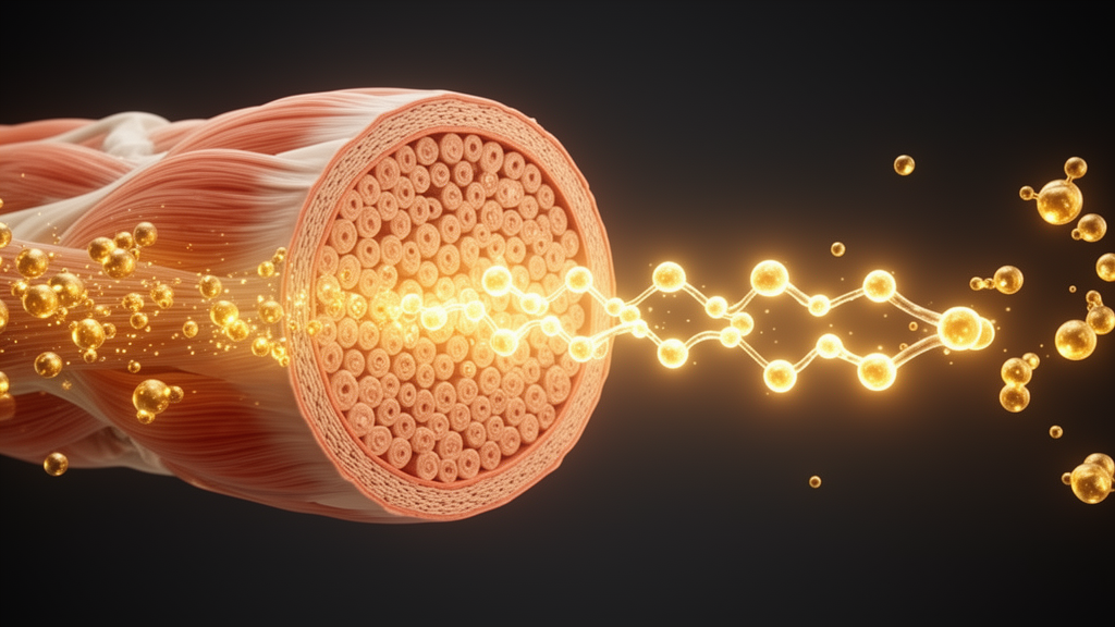
A natural, zero-calorie sweetener derived from stevia has been shown to restore glucose uptake in the skeletal muscle of mice with prediabetes, and researchers have now traced the molecular route it takes to get there. In a study published in Food Production, Processing and Nutrition, a team led by Changfa Zhang and Guowei Li reports that stevioside, a diterpene glycoside widely used as a sugar substitute, improved fasting blood glucose, glucose tolerance, muscle glycogen stores and blood lipid profiles in high-fat diet-fed male mice, and that these benefits depended on the JAK2/STAT3 signaling pathway acting through a downstream effector molecule called IL6ra.
The work focused on the prediabetes stage, the period in which blood glucose abnormalities develop gradually and can still be delayed or prevented by appropriate intervention. Prediabetes is considered a key precursor of type 2 diabetes mellitus, a condition that affected 540 million adults globally in 2021 and is projected to reach roughly 700 million by 2045, with more than 90 percent of cases being type 2 diabetes. Because skeletal muscle accounts for approximately 80 percent of postprandial glucose disposal, the researchers reasoned that targeting glucose metabolism in this tissue could be a worthwhile strategy for preventing progression from prediabetes to full diabetes.
To build the model, the team fed male C57BL/6J mice a high-fat diet deriving 60 percent of energy from fat for 12 weeks, after which the animals had fasting blood glucose levels between 7.0 and 11.1 mmol/L but had not crossed the diabetic threshold above 11.1 mmol/L. The prediabetic mice were then randomly divided into three groups of six animals each. One group served as an untreated prediabetic control, while the other two received aqueous solutions of stevioside at 200 mg/L or 400 mg/L for eight weeks. The lower dose corresponds to the maximum concentration permitted in beverages in the European Union, and the higher dose reflects the maximum suggested by the World Health Organization and the Food and Agriculture Organization. A normal chow diet group provided a healthy baseline.
After eight weeks of treatment, the 400 mg/L stevioside group showed significantly lower fasting blood glucose, plasma insulin and HOMA-IR than the untreated prediabetic group, along with significantly improved glucose tolerance in intraperitoneal glucose tolerance tests. Serum markers of oxidative stress also shifted favorably, with differences in malondialdehyde and catalase levels between the treated and untreated prediabetic groups, and all four measured lipid parameters, including triglycerides, total cholesterol, HDL cholesterol and LDL cholesterol, benefited from the intervention. Notably, the 200 mg/L dose produced significant increases in phosphorylated JAK2 in muscle but did not significantly raise phosphorylated STAT3, suggesting that the stronger metabolic effects were concentrated at the higher dose.
To find the mechanism, the researchers performed RNA sequencing on gastrocnemius muscle from the 400 mg/L treatment group and the untreated prediabetic group. The analysis identified 87 differentially expressed genes, and KEGG pathway enrichment pointed to the JAK/STAT signaling pathway as significantly altered. Within that pathway, the gene encoding IL6ra, the alpha subunit of the interleukin-6 receptor complex, stood out as markedly upregulated after stevioside intervention. Western blot analysis of muscle protein extracts confirmed the pattern: phosphorylated JAK2 was significantly elevated in both treatment groups compared with the untreated prediabetic group, phosphorylated STAT3 was significantly increased at the 400 mg/L dose, and IL6ra protein and mRNA levels, which had been reduced by the high-fat diet, were rescued by treatment.
Histology added a structural dimension to the metabolic findings. Periodic acid-Schiff staining of gastrocnemius sections revealed that the high-fat diet had depleted muscle glycogen, with markedly lighter staining and reduced glycogen deposition, while both stevioside doses restored glycogen content in the muscle fibers. A quantitative glycogen assay confirmed that muscle glycogen levels rose significantly in the 400 mg/L group. Hematoxylin and eosin staining showed no pathological differences between the groups, with no inflammatory cell infiltration, fiber necrosis, degeneration or interstitial edema, indicating that the sweetener did not alter muscle tissue morphology. In cultured C2C12 myotubes, the team further showed that palmitic acid treatment reduced phosphorylated GSK3β at Ser9 and increased phosphorylated GYS1 at Ser641, a combination that suppresses glycogen synthesis, and that stevioside co-treatment reversed both changes, promoting glycogen synthesis through the rate-limiting enzyme of the pathway.
The cellular experiments extended the finding to a controlled setting. Treating differentiated C2C12 myotubes and primary mouse skeletal muscle cells with 0.3 mM palmitic acid for 24 hours impaired glucose uptake, measured with the fluorescent glucose analog 2-NBDG, and stevioside co-treatment at 50 micromolar markedly restored uptake in both cell types while raising phosphorylated JAK2, phosphorylated STAT3 and IL6ra protein levels. To test causality, the researchers used the JAK2 inhibitor ruxolitinib and the STAT3 inhibitor bosutinib. Each inhibitor reduced glucose uptake in the myotubes, and stevioside rescued the inhibition while restoring the suppressed phosphorylation and IL6ra expression. Bosutinib did not affect phosphorylated JAK2, consistent with STAT3 acting downstream of JAK2. When the team silenced IL6ra with shRNA plasmids, glucose uptake fell, but stevioside treatment again improved it, and the knockdown left phosphorylated JAK2 and STAT3 unchanged, supporting IL6ra as a downstream effector rather than an upstream regulator of the pathway.
The authors frame the result as filling a tissue-specific gap in the known hypoglycemic actions of stevioside. Their previous work had shown that the compound alleviated insulin resistance in high-fat diet-induced prediabetic female mice through the hepatic IRS1/PI3K/AKT signaling cascade, but that study did not examine skeletal muscle. The new findings suggest that stevioside acts on distinct signaling pathways in different metabolic organs, and the authors note that interleukin-6 signaling is highly context-dependent, with the function of the JAK2/STAT3 pathway varying by cell type and physiological environment. They also point to prior work linking IL6ra to glucose and energy metabolism, including evidence that IL6ra deficiency leads to severely impaired blood glucose regulation and that IL6ra on skeletal muscle participates in exercise-enhanced glucose uptake and fatty acid oxidation.
Several limitations temper the conclusions. The specific upstream molecular targets of stevioside remain unclear, including whether it directly binds cytokine receptors or influences the activity of related kinases or phosphatases. The in vitro experiments, while supportive, cannot fully capture the complexity of cytokine networks, systemic effects and inter-tissue communication in living animals, and the authors call for long-term studies to clarify the beneficial or potentially harmful roles of interleukin-6 signaling in prediabetes. They also propose incubating gastrocnemius muscle ex vivo with stevioside to measure glucose uptake at the organ level, which would provide more direct evidence than the current design. The study used only male mice, six animals per group, and the 400 mg/L dose drove most of the significant metabolic improvements, leaving the lower, beverage-equivalent dose with a more limited demonstrated effect.
Within those boundaries, the study offers a mechanistic account of how a widely consumed natural sweetener might support muscle glucose handling during the reversible window of prediabetes. The loss-of-function and gain-of-function design, combining pathway inhibitors with targeted gene knockdown, allowed the team to establish the ordering of molecules within the JAK2/STAT3/IL6ra axis rather than merely observing correlations. The authors suggest that stevioside may serve as a dietary strategy to help prevent the progression of type 2 diabetes, while emphasizing that the findings warrant further validation before any translation to human dietary recommendations.
Subject of Research: The effect of stevioside on skeletal muscle glucose uptake in prediabetic mice via the JAK2/STAT3/IL6ra signaling pathway
Article Title: Stevioside enhances skeletal muscle glucose uptake in high-fat diet-induced prediabetic mice via the JAK2/STAT3/IL6ra axis
Article References: Zhang, C., Kang, L., Peng, M., Li, K., Li, Z., Wang, R., Qu, H., & Li, G. (2026). Stevioside enhances skeletal muscle glucose uptake in high-fat diet-induced prediabetic mice via the JAK2/STAT3/IL6ra axis. Food Production, Processing and Nutrition, 8(1), Article 79. https://doi.org/10.1186/s43014-026-00438-0
Image Credits: AI Generated
DOI: 10.1186/s43014-026-00438-0
Keywords: stevioside, prediabetes, skeletal muscle, glucose uptake, JAK2/STAT3, IL6ra, type 2 diabetes, glycogen synthesis, high-fat diet, natural sweetener, mouse study, C2C12 myotubes
News Source: Daisy Hatcher. (October 28, 2026). Stevioside Boosts Muscle Glucose Uptake in Prediabetic Mice Through a Newly Mapped Signaling Axis. Scienmag.