Cisplatin is one of the most widely used and most effective chemotherapy drugs in the world, deployed against testicular, ovarian, bladder, lung and many other cancers. Yet every oncologist knows its shadow side: in a substantial fraction of patients, the drug triggers acute kidney injury, a sudden and sometimes lasting collapse of renal function that can force dose reductions, treatment delays or discontinuation of a potentially curative therapy. For decades, researchers have chipped away at the mechanisms behind this toxicity, implicating oxidative stress, mitochondrial damage and inflammatory cascades in the death of the kidney’s hardest-working cells. A new study published in Cellular and Molecular Life Sciences adds a striking and previously overlooked player to that list: a nuclear splicing factor called LUC7L2, which appears to push kidney cells toward ferroptosis, an iron-dependent form of cell death, by sabotaging the RNA processing of a single critical gene.
The research, led by Bin Fang, Fangfang He and Ling Xie of the Department of Nephrology at Union Hospital, Tongji Medical College, Huazhong University of Science and Technology, together with colleagues in Wuhan, Xining and Shanghai, focuses on the proximal tubular epithelial cells that line the kidney’s filtering tubules. These cells absorb the bulk of cisplatin from the urine, which is precisely why they bear the brunt of the drug’s toxicity. The team hypothesized that the vulnerability of these cells might be governed not only by which genes are switched on or off, but by how their messenger RNA transcripts are spliced, the molecular editing step that removes introns and stitches exons together into a mature, protein-coding message.
At the center of the story is SLC7A11, the gene encoding the light chain of system xc−, an antiporter that imports cystine into the cell in exchange for glutamate. Cystine is the raw material for glutathione, the cell’s most abundant antioxidant, and glutathione in turn fuels glutathione peroxidase 4, or GPX4, the only enzyme capable of neutralizing the lipid peroxides that accumulate during ferroptosis. When SLC7A11 activity falls, the antioxidant buffer thins, lipid membranes oxidize, and the cell ruptures in the characteristic iron-fueled burn of ferroptosis. Because SLC7A11 sits at this choke point, it has become one of the most intensively studied regulators of ferroptosis in cancer biology and, increasingly, in kidney disease.
What makes the new work unusual is its focus on LUC7L2, a component of the spliceosome previously studied mainly in the context of glucose metabolism in cancer cells. The authors began by showing that LUC7L2 expression rises in models of cisplatin-induced acute kidney injury, both in cultured proximal tubule cells and in animal models. They then manipulated the factor in both directions: silencing it with small interfering RNAs, boosting it with plasmid overexpression, and knocking it down in living animals using adeno-associated virus 9 vectors. The functional readouts were the standard arsenal of ferroptosis assays: cell viability measured by CCK-8, lipid peroxidation tracked through markers such as malondialdehyde and 4-hydroxynonenal, iron accumulation, glutathione levels and GPX4 expression.
The results were consistent across every platform. When LUC7L2 was knocked down, ferroptotic damage receded: cells survived better, lipid peroxidation markers fell, glutathione was preserved and GPX4 held steady. When LUC7L2 was overexpressed, the opposite occurred, with kidney cells sliding more readily into ferroptotic death. In vivo, adeno-associated virus-mediated knockdown of LUC7L2 attenuated renal injury in the cisplatin model, as reflected in the study’s biochemical and histological assessments. In other words, LUC7L2 was not a passive bystander in the injured kidney but an active promoter of the damage.
The mechanistic core of the paper lies in how LUC7L2 acts on SLC7A11. Using exon-specific splicing assays and RNA immunoprecipitation, the researchers showed that the splicing factor binds directly to SLC7A11 pre-messenger RNA and is associated with increased exon skipping. Skipping events that introduce a premature termination codon produce non-productive transcripts, messages that are either degraded or translated into truncated, useless proteins. Under cisplatin stress, LUC7L2 was associated with precisely this kind of non-productive splicing, leaving cells with less functional SLC7A11 protein, diminished cystine import, depleted glutathione and, ultimately, heightened susceptibility to ferroptosis. This is a post-transcriptional layer of control, operating after the gene has been transcribed, that complements rather than duplicates the better-known transcriptional regulation of SLC7A11.
That transcriptional layer, the study shows, belongs to p53, the famous tumor-suppressor protein that cisplatin also activates. The authors describe a dual-pathway mechanism converging on SLC7A11: cisplatin-activated p53 represses transcription of the gene, while LUC7L2 simultaneously corrupts the splicing of whatever pre-messenger RNA is still produced. The two arms of the attack squeeze the antioxidant system from both ends, one throttling the gene’s output at the source and the other degrading the quality of what remains. When the team used pifithrin-alpha, a p53 inhibitor, in their experiments, the transcriptional arm of this squeeze was relieved, underscoring how the two mechanisms operate in parallel rather than in sequence.
To establish that the phenomenon is relevant beyond cell culture and rodents, the researchers reanalyzed public single-cell RNA sequencing datasets to map where LUC7L2 and SLC7A11 are expressed across the many cell types of the kidney, and then validated their findings in human kidney biopsy specimens and urine samples from patients. The single-cell analysis helped define the cell-type-specific expression patterns of the regulatory axis, pointing to the proximal tubule as the site where the LUC7L2–SLC7A11 interaction matters most. The human validation is particularly significant for a field in which promising animal results frequently fail to translate, and it suggests that the splicing factor’s expression could serve as a biomarker of ferroptotic activity in injured kidneys.
The clinical implications run in two directions. As a biomarker, LUC7L2 offers a potential early signal of ferroptotic kidney damage, detectable in urine, that could help clinicians identify patients at risk before blood urea nitrogen and creatinine rise, which are late indicators that appear only after substantial nephron loss has already occurred. As a therapeutic target, the study suggests that damping LUC7L2 activity, or otherwise protecting SLC7A11 splicing, could shield the kidney during cisplatin treatment without necessarily interfering with the drug’s anti-tumor effect, since the protective pathway operates in host tissue rather than in the cancer cells themselves. Ferroptosis inhibitors such as ferrostatin-1 and liproxstatin-1, which the authors used as experimental tools, and antioxidants like N-acetylcysteine represent existing pharmacological handles on the downstream pathway, but a splicing-level intervention would act further upstream and with greater specificity.
There are, of course, caveats. Splicing factors participate in the maturation of thousands of transcripts, and any therapeutic strategy aimed at LUC7L2 would need to account for its broader portfolio of substrates, particularly given its documented role in cancer cell metabolism. The study, which was supported by the National Natural Science Foundation of China and other national and provincial programs, also relies on models of one specific cause of acute kidney injury; whether the same LUC7L2–SLC7A11 axis operates in ischemic, septic or contrast-induced kidney injury remains to be tested. Even so, the work opens a genuinely new chapter in renal protection research. It demonstrates that the fate of a cell under chemotherapy stress can hinge not just on gene expression but on gene editing at the RNA level, and it identifies a concrete, druggable-looking node where ferroptosis, splicing and p53 signaling intersect. For the millions of patients who receive cisplatin each year, that intersection may one day mark the difference between a kidney that recovers and one that does not.
Subject of Research: The role of the splicing factor LUC7L2 in regulating SLC7A11-dependent ferroptosis during cisplatin-induced acute kidney injury
Article Title: LUC7L2 promotes ferroptosis via alternative splicing of SLC7A11 in cisplatin-induced acute kidney injury
Article References: Fang, B., He, F., Xie, L., Yuan, Q., Ye, C., Song, A., Wang, X., Xiong, W., Su, H., Luo, P., Zhang, X., & Zhang, C. (2026). LUC7L2 promotes ferroptosis via alternative splicing of SLC7A11 in cisplatin-induced acute kidney injury. Cellular and Molecular Life Sciences. https://doi.org/10.1007/s00018-026-06411-x
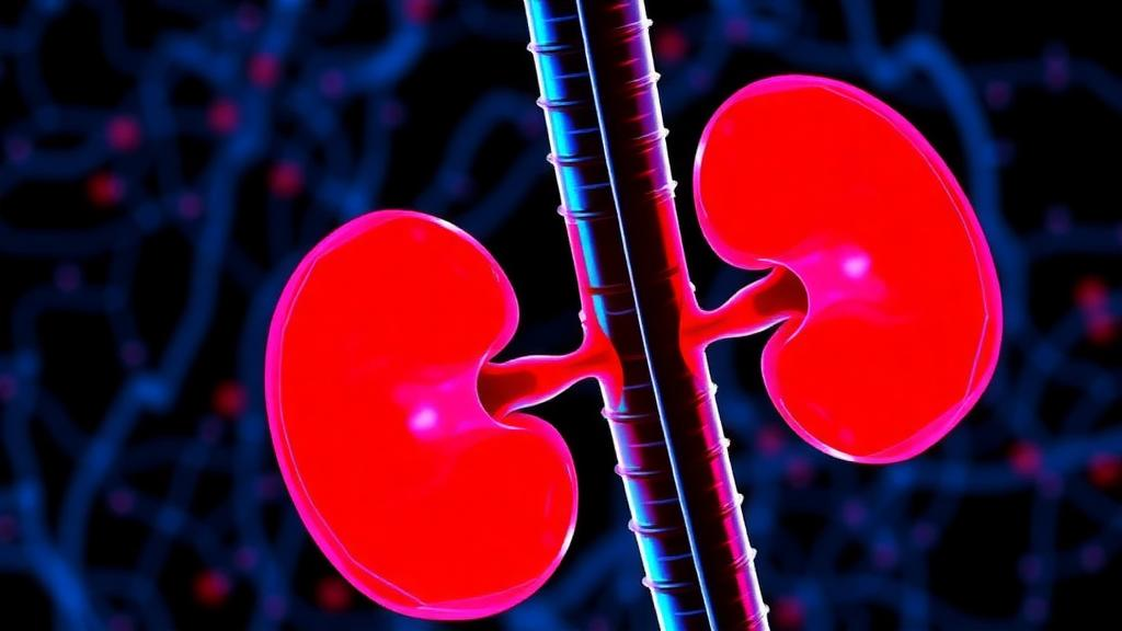
Image Credits: AI Generated
DOI: 10.1007/s00018-026-06411-x
Keywords: LUC7L2, SLC7A11, ferroptosis, acute kidney injury, cisplatin, alternative splicing, p53, GPX4, glutathione, proximal tubular cells, lipid peroxidation, single-cell RNA sequencing
News Source: Nathaniel Bowman. (October 6, 2026). Splicing Factor LUC7L2 Emerges as a Driver of Kidney Damage from Chemotherapy. Scienmag.