Spatial single-cell profiling of human kidney tissue has revealed that acute interstitial nephritis, a severe cause of acute kidney injury affecting fifteen to twenty percent of biopsy-confirmed cases, is organized by distinct molecular circuits and inflammatory microenvironments that distinguish it from non-immune kidney damage. Investigating biopsy specimens using high-resolution imaging mass cytometry alongside single-cell spatial transcriptomics, researchers mapped how injured epithelial cells in proximal tubules communicate directly with infiltrating immune cells. Acute interstitial nephritis frequently arises from drug reactions, notably including modern cancer immune checkpoint inhibitors, yet therapeutic options remain largely restricted to discontinuation of triggering medications and empirical corticosteroid regimens that fail in twenty to forty percent of patients. By comparing kidney biopsies from patients with acute interstitial nephritis against biopsies exhibiting acute tubular injury and healthy reference kidney tissues, the investigators uncovered how structural epithelial cells actively drive immune recruitment through local complement production, metabolic reprogramming, and self-amplifying interferon-driven chemokine networks.
The investigation encompassed one hundred and six kidney tissue specimens gathered across two independent clinical medical centers, comprising a discovery cohort of sixty-five specimens from Yale and a validation cohort of forty-one specimens from Johns Hopkins University. Among these samples, thirty-seven represented pathologist-adjudicated acute interstitial nephritis, thirty-seven represented acute tubular injury, and thirty-two served as histopathologically evaluated reference tissues obtained from living donor transplant biopsies or tumor-remote nephrectomy margins. The imaging mass cytometry panel employed thirty-one metal-conjugated antibodies targeting structural nephron markers, immune cell lineages, and functional activation states, generating single-cell spatial proteomic profiles across more than two and a half million cells. To achieve deeper transcriptomic characterization, the team performed single-cell spatial transcriptomics on twenty tissue samples from the discovery cohort using a platform combining more than five thousand human pan-tissue genes with a custom one-hundred-gene panel dedicated to renal tubule segments, immune markers, and fibrosis pathways.
Through unsupervised clustering of imaging mass cytometry proteomic data, the researchers delineated four distinct states across cortical proximal tubule cells: healthy epithelium, dedifferentiated tubules, injured state one, and injured state two. While healthy proximal tubule cells expressed abundant megalin and aquaporin-one, injured state one cells exhibited loss of apical brush borders and expression of kidney injury molecule-one without vascular cell adhesion molecule-one. In contrast, injured state two cells demonstrated prominent vascular cell adhesion molecule-one expression, cellular flattening, dilated tubular lumens, and adjacent peritubular scarring. Blinded histological evaluation confirmed that composite injury scores increased along this continuum from healthy tissue to injured state one. Crucially, while overall proximal tubule cell density declined in acute interstitial nephritis compared with reference kidneys, vascular cell adhesion molecule-one positive injured state two cells expanded dramatically, constituting twenty-eight point nine percent of proximal tubules in nephritis compared to five point one percent in acute tubular injury.
This pronounced expansion corresponded to a six-and-a-half-fold elevation in vascular cell adhesion molecule-one positive tubule density in acute interstitial nephritis relative to acute tubular injury, reaching a median of two hundred and sixty-seven cells per square millimeter compared to forty-one cells per square millimeter. Conversely, the density of injured state one cells showed no significant statistical difference between acute interstitial nephritis and acute tubular injury, indicating that early tubular injury occurs across diverse renal insults, whereas the accumulation of vascular cell adhesion molecule-one positive tubules represents a pathological feature characteristic of interstitial nephritis. Spatial neighborhood analysis demonstrated that these distinct epithelial injury states inhabited markedly divergent cellular surroundings. Healthy proximal tubules were bordered predominantly by microvascular endothelial cells, whereas injured state one tubules were enriched for adjacent mononuclear phagocytes, and vascular cell adhesion molecule-one positive injured state two tubules showed selective spatial association with infiltrating T lymphocytes and organized lymphoid clusters.
Spatial transcriptomic analysis established that injured proximal tubules in acute interstitial nephritis serve as primary regional sources of complement components, directly linking structural epithelial damage to local innate immune recruitment. Cortical tubular epithelial cells accounted for sixty-three point seven percent of total complement C3 messenger RNA in nephritis tissue, far exceeding the twenty-one point nine percent contributed by infiltrating immune cells. Proximal tubules represented the dominant epithelial source, generating seventy-eight point two percent of all tubular complement C3 transcripts. Cellular C3 expression escalated progressively along the tubular injury trajectory, rising from uninjured cells to kidney injury molecule-one positive cells and reaching highest levels within vascular cell adhesion molecule-one positive tubules. A parallel upregulation occurred in thick ascending limb cells that had lost normal uromodulin expression, where complement C3 transcripts were twenty-three times higher than in healthy thick ascending limbs and more than three times higher than in dedifferentiated limbs from acute tubular injury.
Tissue-level tubular complement production strongly correlated with the spatial abundance of mononuclear phagocytes expressing the C3a receptor, known as C3AR1. Spatially constrained modeling within a twenty-micrometer radius demonstrated that predicted paracrine C3 to C3AR1 ligand-receptor interactions were nearly fivefold higher in acute interstitial nephritis than in reference kidneys and fifty percent higher than in acute tubular injury. The primary recipient cells receiving these epithelial complement signals were pro-inflammatory macrophages co-expressing CD74 and high levels of lysosomal acid lipase, which accounted for fifty percent of all predicted tubule-to-macrophage complement interactions. This specific macrophage recruitment pattern was distinct from a second pro-inflammatory macrophage subset marked by CXCL9 and CXCL10 expression, which showed elevated tissue density in acute interstitial nephritis but did not concentrate at tubular basolateral surfaces, demonstrating that renal epithelial cells deploy complement gradients to recruit discrete innate immune subsets directly to vulnerable tubule-interstitial interfaces.
Applying a geometric deep learning framework called SORBET to identify transcripts within neighboring cellular niches predictive of tubular epithelial injury states revealed nicotinamide phosphoribosyltransferase, or NAMPT, as the strongest discriminator of vascular cell adhesion molecule-one positive microenvironments. NAMPT serves both as an essential rate-limiting enzyme in intracellular nicotinamide adenine dinucleotide biosynthesis and as an extracellular inflammatory cytokine. In acute interstitial nephritis cortex, NAMPT expression increased in a stepwise fashion across surrounding non-tubular cells as the local density of adjacent vascular cell adhesion molecule-one positive tubules grew. Proximal tubule cells themselves also showed stepwise NAMPT upregulation, increasing from eight point six percent in baseline tubules to seventeen point three percent in vascular cell adhesion molecule-one positive tubules. This coordination between damaged epithelial cells and infiltrating immune cells points to a localized metabolic-inflammatory niche in which compromised cellular energy pathways align with intense local inflammatory responses.
The immune infiltrate in acute interstitial nephritis was quantitatively dominated by T lymphocytes, whose cortical median density was more than fivefold greater than that observed in acute tubular injury. Single-cell spatial transcriptomics demonstrated that CD8-positive cytotoxic T cells constituted sixty-two percent of this lymphocytic infiltrate, with interferon-activated T cell subsets displaying the steepest relative expansions over acute tubular injury. Spatial ligand-receptor interaction modeling identified an extraordinary forty-four-fold enrichment of predicted paracrine CXCL9 to CXCR3 signaling interactions in nephritis compared to tubular injury, with a median density of fifty-two point one interactions per square millimeter versus one point two. Rather than occurring between epithelial cells and leukocytes, these interactions were overwhelmingly dominated by homotypic T cell-to-T cell amplification networks, where CD8-positive T cell pairs comprised forty-one point two percent of all CXCL9-CXCR3 signaling pairs, driven substantially by interferon-activated and follicular cytotoxic T cell subpopulations.
These potent chemokine circuits operated in an environment characterized by a striking depletion of regulatory immune checkpoints. In healthy reference kidney tissues, the investigators routinely detected tissue-resident memory CD8-positive T cells co-expressing inhibitory receptors including LAG3, TIGIT, and programmed cell death protein-one. However, these checkpoint-high regulatory cells were completely absent in seven out of eight patients with acute interstitial nephritis, and their absence directly tracked with elevated CXCL9-CXCR3 signaling intensity. Furthermore, the signaling networks concentrated within ectopic lymphoid aggregates, which were identified in seven out of eight nephritis patients but only two of seven acute tubular injury patients and zero reference kidneys. Within these aggregates, CXCL9-CXCR3 interaction densities were more than three times higher than in surrounding cortex. Although these aggregates harbored B cells, T cells, and dendritic cells alongside organizing chemokines CCL19 and CCL21, they lacked classical lymphoid stromal elements like follicular dendritic cells.
The researchers also uncovered a striking divergence regarding macrophage prognostic value: higher density of CD206-negative pro-inflammatory macrophages correlated with improved six-month kidney function recovery in steroid-treated nephritis patients, whereas macrophage density in acute tubular injury correlated negatively with functional recovery. In interpreting these findings, the authors highlighted important methodological boundaries, noting that cross-sectional biopsy profiling cannot establish directional causality between tubular phenotypic shifts and immune activation, nor can it track dynamic cellular progression over time. In addition, seventy-seven percent of nephritis patients received post-biopsy corticosteroids, which may confound outcome correlations evaluated across modest patient subsets. Because the spatial transcriptomic cohort included two reference cortical samples, statistical contrasts were formally conducted between nephritis and tubular injury. Nevertheless, identifying the coordinated triad of proximal tubule complement synthesis, metabolic NAMPT signaling, and lymphoid CXCL9-CXCR3 amplification loops nominates specific targeted therapeutic avenues beyond generalized immunosuppression.
Subject of Research: Spatial single-cell multiomic analysis of inflammatory microenvironments in human acute interstitial nephritis
Article Title: Spatial analysis reveals cellular microenvironments and mechanisms of inflammation and injury in acute interstitial nephritis
Article References: Baker, M. L., Kakade, V. R., Budiman, T., Weiß, M., Cunningham, J. M., Sadarangani, S., Lerner, G., Moeckel, G., Rosenberg, A. Z., Parikh, C. R., Kluger, Y., Moledina, D. G., & Cantley, L. G. (2026). Spatial analysis reveals cellular microenvironments and mechanisms of inflammation and injury in acute interstitial nephritis. Nature Communications, 17(1), Article 10262. https://doi.org/10.1038/s41467-026-77210-y
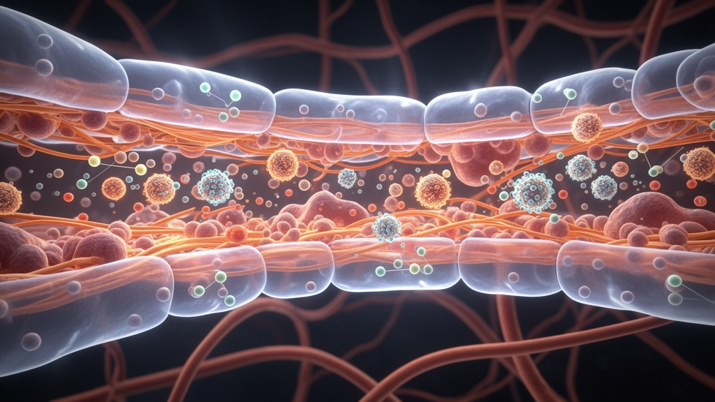
Image Credits: AI Generated
DOI: 10.1038/s41467-026-77210-y
Keywords: acute interstitial nephritis, acute tubular injury, imaging mass cytometry, spatial transcriptomics, proximal tubules, VCAM1, complement C3, C3AR1, NAMPT, CXCL9, CXCR3, CD8+ T cells, tertiary lymphoid structures, interferon-gamma signaling, macrophage polarization, CD206, renal pathology
News Source: Ophelia Keating. (November 21, 2026). Spatial Profiling Reveals Complement and Interferon Circuits Driving Acute Interstitial Nephritis. Scienmag.