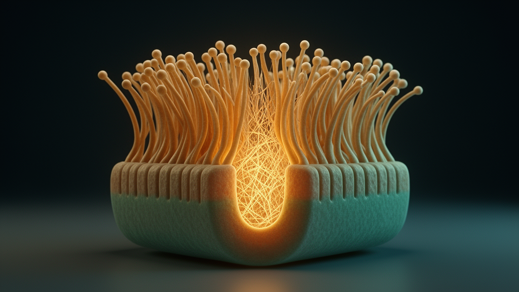
Hearing depends on a structure that most people never think about: the tiny, staircase-shaped bundles of stereocilia that crown the sensory hair cells of the inner ear. Each stereocilium is built around a rigid core of parallel actin filaments, and this core must survive a lifetime of mechanical bending without significant turnover. A new study published in EMBO Reports identifies the transcription factor Sox3 as a central guardian of that architecture, showing in mice that loss of Sox3 causes the stereocilia core to progressively disassemble, while boosting its expression protects the ear against noise-induced damage.
The research team, led by Wen-Xiao Wang, Ling-Yun Zhou and Ting-Ting Du, found that in mice lacking Sox3, the actin cores of stereocilia develop phalloidin-negative gaps, a hallmark of core damage visible under confocal microscopy. These gaps first appeared at about four weeks of age and accumulated with time, ultimately producing hearing loss across the full frequency range and impaired mechanotransduction, the process by which hair cells convert mechanical deflection into electrical signals. Transmission electron microscopy of utricle hair cells confirmed the breakdown, revealing that the normally dense, ordered packing of actin filaments was replaced by disorganized structures interspersed with electron-lucent gaps.
Sox3 belongs to the SoxB1 subgroup of high mobility group box transcription factors, a family that also includes Sox1 and Sox2 and is best known for governing embryonic development and cell fate decisions. Previous work had already connected Sox3 to the auditory system: it is essential for inner ear neurogenesis in chick and zebrafish models, and human genetic studies have associated SOX3 mutations with hearing loss. What the new study adds is a role that extends far beyond development, into the lifelong structural maintenance of the mature hair cell.
Because Sox3-null mice show broad abnormalities, including craniofacial malformations and hypothalamo-pituitary defects, the researchers generated conditional knockouts to isolate the cochlear effect. An otic vesicle-specific deletion, driven by an improved Foxg1-IRES-Cre line, removed Sox3 from the inner ear early in development without the systemic confounds. A second model, crossing Sox3-floxed mice with Atoh1-Cre, deleted the gene specifically in hair cells while sparing supporting cells. Both strategies produced the same picture: normal hair bundle formation during development, followed by progressive actin core deterioration beginning around P30.
The functional consequences were measurable and progressive. Auditory brainstem response recordings showed elevated thresholds in Sox3-deficient mice at P30, worsening with age; by four months, threshold elevations of roughly 20 to 40 dB were present across all tested frequencies. Distortion product otoacoustic emission measurements paralleled the ABR shifts, indicating compromised outer hair cell amplification. At four months, the researchers also used gentamicin uptake as a proxy for mechanotransduction and found markedly reduced uptake in outer hair cells of mutant mice, consistent with the idea that a weakened actin core diminishes hair bundle stiffness and disrupts signal transduction.
Notably, vestibular function told a different story. Utricular hair cells in the knockout mice accumulated the same phalloidin-negative gaps over time, yet vestibular evoked potentials recorded at one and four months showed no significant differences from controls.
The most striking results emerged when the researchers exposed the mice to noise. In P20 animals, before any spontaneous gaps were visible, a two-hour exposure to broadband noise at 117 dB SPL dramatically accelerated gap formation in Sox3-deficient mice compared with controls. In P30 mice exposed to 120 dB SPL for three hours, both groups formed gaps, but the mutants suffered more extensive damage, with widespread stereocilia breakage confirmed by both fluorescence staining and scanning electron microscopy. After a permanent threshold shift-inducing noise exposure, control mice recovered substantially better hearing by seven days, while the mutants did not.
Gap repair itself proved defective. Normally, noise-induced gaps in stereocilia are repaired within about a week, with monomeric gamma-actin accumulating at lesion sites to rebuild the core. In control mice exposed to 117 dB broadband noise, gap counts rose transiently at one hour and returned to baseline by 24 hours. In the Sox3-deficient mice, the initial increase was similar, but the gaps persisted at 24 hours, indicating impaired repair. Interestingly, gamma-actin recruitment to the gaps was unaffected; line scans showed roughly a threefold enrichment of gamma-actin fluorescence at gap centers in both groups, suggesting that Sox3 acts upstream of the recruitment step rather than blocking it.
Overexpression experiments flipped the phenotype. The team engineered knock-in mice carrying an IRES-Sox3 cassette downstream of the endogenous Atoh1 gene, producing hair cell-specific Sox3 overexpression at approximately twofold mRNA levels in heterozygotes. These animals had normal hearing and hair cell counts, but after noise exposure they formed significantly fewer gaps and breaks than controls, with homozygous overexpressors protected more than heterozygotes, a clear gene dosage effect. Under noise conditions that induce permanent threshold shifts, overexpressing mice developed smaller initial threshold shifts and recovered better hearing across the 11 to 22 kHz range over the following two weeks. A genetic rescue experiment, crossing the overexpression allele into the otic vesicle knockout background, substantially reduced gap formation and improved auditory thresholds, confirming specificity.
Mechanistically, RNA sequencing of fluorescently sorted P2 hair cells revealed 354 differentially expressed genes in the absence of Sox3, about a third of them downregulated. Gene ontology analysis pointed strongly toward ribosomal and translational machinery, but among the downregulated genes were the beta-thymosins, particularly Tmsb10, an actin monomer-binding protein that regulates the availability of G-actin for filament assembly. Tmsb4x, the most abundant family member, was also significantly reduced. A conditional Tmsb10 knockout in the otic vesicle reproduced the stereocilia core damage, though in a milder form than Sox3 loss, possibly because Tmsb4x, roughly 150-fold more abundant in hair cells, partially compensates. Tmsb10 knockout mice likewise developed mild hearing loss by P60. In cell culture, siRNA knockdown of TMSB10 or TMSB4X in HeLa and HEI-OC1 cells disrupted actin stress fibers, reduced lamellipodial expansion and caused gamma-actin to mislocalize into peripheral patches, underscoring the protein’s role in organizing actin-based architecture. The authors propose that Sox3, expressed transiently in early postnatal hair cells, may establish an epigenetic landscape that sustains Tmsb10 expression into adulthood, and they identify the Sox3-Tmsb10 axis as a promising target for future therapies against noise-induced hearing loss, while cautioning that the precise chromatin mechanisms and the full downstream network remain to be worked out.
Subject of Research: The role of the transcription factor Sox3 in maintaining the actin core of inner ear hair cell stereocilia and protecting against noise-induced hearing loss in mice
Article Title: Sox3 stabilizes the actin core of hair cell stereocilia essential for hearing
Article References: Wang, W.-X., Zhou, L.-Y., Zhu, Y., Wu, H., & Du, T.-T. (2026). Sox3 stabilizes the actin core of hair cell stereocilia essential for hearing. EMBO Reports. https://doi.org/10.1038/s44319-026-00943-w
Image Credits: AI Generated
DOI: 10.1038/s44319-026-00943-w
Keywords: Sox3, hair cells, stereocilia, actin core, hearing loss, noise-induced hearing loss, mechanotransduction, Tmsb10, beta-thymosin, gap repair, cochlea, mouse models
News Source: Drew Townsend. (October 28, 2026). Sox3 Keeps the Actin Core of Hair Cell Stereocilia Stable for Lifelong Hearing. Scienmag.