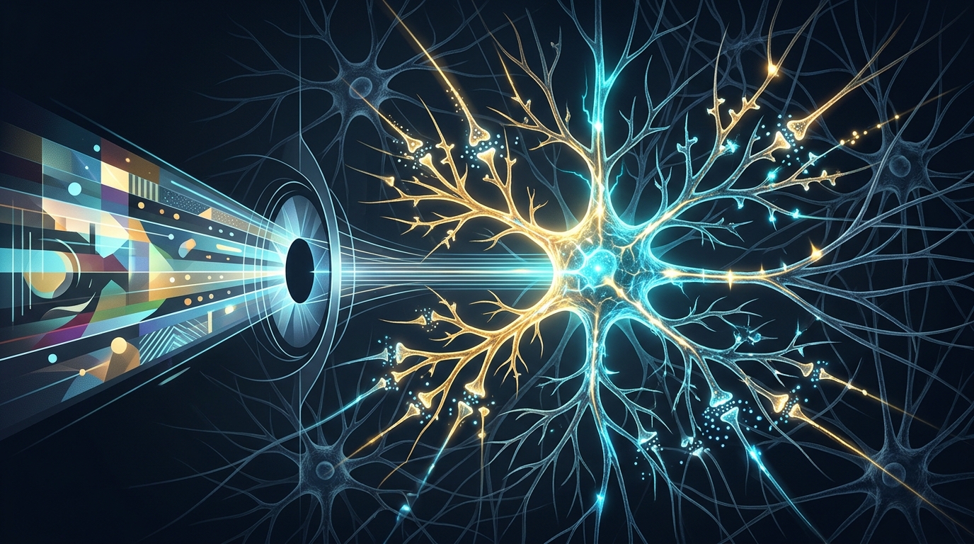
A few glances at a two-tone image can transform meaningless black-and-white blotches into instantly recognizable objects, and researchers have now watched that transformation unfold neuron by neuron in the human brain. In a study published in Nature Neuroscience, a team recorded the electrical activity of 1,104 individual neurons in people with epilepsy while the participants learned to recognize degraded pictures known as Mooney images. The recordings show that after only one or a few exposures to the intact version of a picture, neurons in the occipital cortex and the medial temporal lobe reshaped their firing patterns so that the previously meaningless two-tone image evoked activity resembling the response to the original photograph.
The participants were 13 adult patients with drug-resistant epilepsy who had been implanted with hybrid macro- and microelectrodes at the Freiburg Epilepsy Center in Germany to monitor seizures. Electrode placement was determined solely by clinical need, giving the researchers a rare window onto single-neuron activity in the medial occipital cortex, the parahippocampal cortex, the entorhinal cortex, the hippocampus and the amygdala across 34 recording sessions. The recorded neurons included 163 in medial occipital cortex, 212 in parahippocampal cortex, 49 in entorhinal cortex, 343 in hippocampus and 337 in amygdala. Because the patients volunteered and gave informed consent, the team could sample spikes from individual cells while the participants performed a recognition task that would be impossible to run in typical laboratory animals at this scale.
The task itself exploited the peculiar psychology of Mooney images, which are two-tone black-and-white pictures that most people cannot decode until they have seen the corresponding grayscale photograph. Each trial presented an image for 500 milliseconds followed by a blank screen with a fixation cross for 1,300 milliseconds, and participants pressed a button to report whether they recognized the depicted object. The experiment cycled through three conditions: pre-learning Mooney images shown before any exposure to the intact picture, grayscale originals, and post-learning Mooney images shown after brief learning sequences in which the grayscale version was immediately followed by its two-tone counterpart. Recognition of Mooney images rose from an average accuracy of 4 percent before learning to 83 percent after learning, while grayscale images were recognized at 93 percent accuracy. On average, participants needed only 1.8 learning sequences to master a new image, and the first recognition was confirmed by a verbal report of the object’s identity.
Behavioral timing told a second story. Recognized Mooney images took longer to judge than grayscale photographs, with median response times of 0.53 seconds versus 0.48 seconds, while unrecognized pre-learning Mooney images elicited the slowest responses of all at a median of 0.60 seconds. The researchers interpret the extra time as a sign that interpreting the learned two-tone images demanded additional cognitive effort. Eye-tracking in a separate cohort of ten healthy participants, who showed the same recognition pattern, helped rule out an obvious confound: in roughly 83 percent of trials participants made no saccades during image presentation, and blink rates were negligible, indicating that the images were perceived in a single glance rather than assembled through scanning. Control trials pairing a grayscale image with a mismatched Mooney image produced no learning, with accuracy on the unrelated Mooney images averaging just 7 percent, showing that simple association could not explain the effect.
Among the 1,104 recorded neurons, 318 were classified as visually responsive, meaning their firing rates changed significantly during image presentation compared with baseline. These responsive cells were unevenly distributed: 105 of 163 occipital neurons, 90 of 212 parahippocampal neurons, and 123 across the hippocampus, amygdala and entorhinal cortex, which the team analyzed together as the medial temporal lobe. Response latencies increased along this axis, with occipital neurons firing first at a median of 155 milliseconds after stimulus onset, parahippocampal neurons at 161 milliseconds, and medial temporal neurons at 322 milliseconds. Selectivity followed the opposite gradient, with medial temporal neurons showing the highest stimulus selectivity index at 0.67 compared with 0.56 in both occipital and parahippocampal cortex, while occipital neurons carried the strongest signal-to-noise ratio at 8.9. The authors describe this as a hierarchical organization in which latencies and selectivity rise as signals flow from occipital cortex toward the medial temporal lobe.
The most striking results emerged when the researchers compared responses to the same Mooney image before and after learning. Although the pixels were identical in both conditions, firing patterns changed dramatically. A hippocampal neuron that was silent to all Mooney images before learning responded selectively to one specific Mooney image afterward, mirroring its response to the grayscale original. To quantify this at the population level, the team defined each neuron’s preferred and nonpreferred images from grayscale responses alone and then tracked responses to those same images in the Mooney conditions. Occipital neurons showed little selectivity for Mooney images before learning, but after learning, responses to preferred images significantly exceeded responses to nonpreferred ones, with the difference emerging 300 to 350 milliseconds after image onset, much later than the 100 to 150 milliseconds seen for grayscale images. Medial temporal neurons showed the same pattern, with selectivity appearing at 350 to 400 milliseconds after learning, and parahippocampal neurons showed no reliable selectivity change in either Mooney condition.
To test whether these changes made image identity genuinely readable from brain activity, the researchers trained linear support vector machine decoders to predict which of three images a participant was viewing from population firing patterns, using pseudopopulations of neurons pooled across participants. Grayscale images were decoded best in all regions, reaching 90 percent accuracy in occipital cortex, 93 percent in parahippocampal cortex and 76 percent in the medial temporal lobe against a 33 percent chance level. For Mooney images the regions diverged: occipital neurons decoded image identity both before and after learning, improving from 62 percent to 80 percent accuracy, whereas parahippocampal and medial temporal neurons decoded at chance before learning but reached 63 percent and 57 percent accuracy, respectively, after learning. Reliable decoding emerged during image presentation and briefly persisted after stimulus offset before returning to chance. Control analyses showed that slow firing-rate drifts over the session could not account for these results, since nonresponsive neurons supported no decoding and the findings held after excluding neurons with the largest firing-rate trends.
Cross-condition analyses then revealed the temporal structure of the learning effect. When classifiers trained on grayscale responses were tested on post-learning Mooney responses, decoding succeeded in both occipital cortex and medial temporal lobe, reaching up to 62 percent and 50 percent accuracy respectively, but failed for pre-learning Mooney images in the medial temporal lobe. The generalization matrices were asymmetric: grayscale response patterns at 175 milliseconds most closely resembled post-learning Mooney patterns at 300 milliseconds in occipital cortex, a 125-millisecond offset, while in the medial temporal lobe grayscale patterns at 325 milliseconds matched learned Mooney patterns at 425 milliseconds, a 100-millisecond offset. In other words, with extra processing time, the brain’s response to a learned two-tone image evolves to resemble its response to the intact photograph. Consistent with this, individual medial temporal neurons responded 110 milliseconds later to learned Mooney images than to grayscale images, whereas occipital latencies did not differ between conditions.
The authors argue that these dynamics point to occipital cortex, and potentially other neocortical areas, as the engine of rapid perceptual learning, with the medial temporal lobe arriving late to the computation rather than driving it. Because the generalization delays in the two regions were comparable, they suggest the temporal lag observed in occipital cortex may simply propagate downstream, and they explicitly note that direct causality was not established in this study. The interpretation aligns with earlier work showing that memory-impaired patients with medial temporal lobe lesions can still perform one-trial perceptual learning tasks, hinting that this form of fast learning may not require the memory system traditionally credited with storing new information. The team also connects the delayed decoding to recurrent processing, in which signals circulate between visual areas and higher-order cortical regions until a degraded image resolves into an object.
Beyond its implications for neuroscience, the study offers a biological benchmark for artificial intelligence systems, which typically need vast supervised datasets to learn object recognition, whereas humans achieve few-shot learning from one or two exposures. The researchers suggest that recognizing Mooney images relies on learning figure-ground segregation and image completion rather than expanding tuning to entirely novel objects, and they frame their single-neuron measurements as constraints for computational models that incorporate recurrent computations for rapid learning. The data and code from the study are publicly available, and the findings sketch a picture of perception as an active reconstruction: within roughly 300 milliseconds in occipital cortex and 425 milliseconds in the medial temporal lobe, newly learned knowledge is woven into the visual response itself, turning noise into meaning.
Subject of Research: Single-neuron mechanisms of rapid visual perceptual learning in the human brain
Article Title: Single-neuron encoding of rapidly learned visual information reshapes human perception
Article References: Armendariz, M., Blumberg, J., Singer, J., Aiple, F., Kim, J., Domínguez-Iturza, N., Brandt, A., Reinacher, P., Schulze-Bonhage, A., & Kreiman, G. (2026). Single-neuron encoding of rapidly learned visual information reshapes human perception. Nature Neuroscience. https://doi.org/10.1038/s41593-026-02496-6
Image Credits: AI Generated
DOI: 10.1038/s41593-026-02496-6
Keywords: Mooney images, single-neuron recordings, occipital cortex, medial temporal lobe, visual recognition, rapid learning, epilepsy patients, neuronal decoding, perceptual learning, recurrent processing, Single-neuron, encoding
News Source: Cassandra Pierce. (November 4, 2026). Single Neurons Reveal How the Brain Learns to See in Seconds. Scienmag.