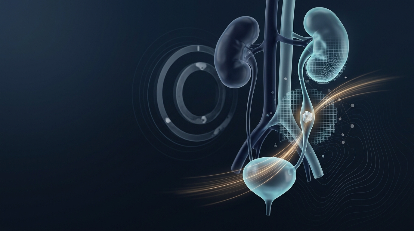
Kidney stones lodged in the ureter force an uncomfortable decision: wait and hope the stone passes on its own, or intervene early with surgery. Current guidance leans heavily on stone size, stone location and clinical judgement, yet these factors leave considerable uncertainty about which patients will actually succeed with conservative management. A new retrospective cohort study, published in BMC Urology, reports that a machine learning model combining radiomic features extracted from non-contrast CT scans with routine clinical variables can estimate the probability of spontaneous stone passage within four weeks with substantially greater accuracy than clinical information alone.
The research team, led by Fan-jie Wei, Yu-song Weng, Shi-ping Zeng, Xin-tao Tan and Jun Huang, retrospectively analysed 274 patients with solitary ureteral stones measuring 10 millimetres or smaller who underwent conservative treatment between January 2021 and August 2025. Time zero was defined as the date of the baseline CT scan at which conservative management was decided, and patients were grouped according to whether the stone passed within four weeks. From manually segmented stone regions on the scans, the investigators extracted 851 radiomic features, quantitative descriptors of image texture and shape that are invisible to the naked eye but statistically informative.
Feature selection proceeded in stages designed to guard against overfitting. Reproducible and non-redundant features were screened by observer-agreement testing, Spearman correlation analysis and least absolute shrinkage and selection operator regression, a technique that shrinks coefficients toward zero to eliminate features that add little predictive information. Eight radiomic features survived this pipeline. The researchers then built three families of models, using clinical variables alone, radiomics alone, and both combined, with four different algorithms: elastic net regression, support vector machine, random forest and LightGBM. Hyperparameters were tuned by 5-fold cross-validation, and final performance was assessed by bootstrap internal validation, calibration analysis, decision curve analysis and the DeLong test for comparing correlated receiver operating characteristic curves.
The headline result concerns the combined random forest model. In bootstrap validation it achieved an area under the curve of 0.928, with a 95% confidence interval of 0.896 to 0.959. That figure was statistically higher than the clinical LightGBM model, which reached an AUC of 0.837 (95% CI 0.789 to 0.884, P < 0.001), and higher than the radiomics random forest model at 0.856 (95% CI 0.809 to 0.903, P = 0.029). An AUC of 0.928 means the model ranked a randomly chosen patient who passed their stone above a randomly chosen patient who did not about 93% of the time, a level of discrimination that, if confirmed externally, would mark a meaningful advance over the conventional size-and-location heuristic.
Discrimination alone is not enough for a predictive tool, so the team examined calibration as well. The combined model showed acceptable calibration, with a Hosmer–Lemeshow P value of 0.591, indicating no statistically significant mismatch between predicted probabilities and observed passage rates. Decision curve analysis found net benefit across threshold probabilities from 5% to 99%, suggesting the model could in principle support decisions across a wide range of risk tolerances. SHAP analysis, a technique that attributes model output to individual inputs, identified CT texture features and ureteral wall thickness as important contributors to prediction, hinting that the model may be capturing genuine anatomical and tissue signals rather than statistical noise.
Because some patients initially assigned to conservative care eventually cross over to intervention, the researchers performed sensitivity analyses. When the 18 crossovers were counted as non-passage, the combined model still yielded an AUC of 0.914 (95% CI 0.878 to 0.949). An exploratory subgroup analysis focused on the intermediate stone-size category, examining 55 patients with 5 to 8 millimetre stones in the proximal or middle ureter, where the decision to wait or operate is most contested. In that subgroup the combined model achieved an AUC of 0.881 (95% CI 0.782 to 0.979), although the wide confidence interval reflects the small sample size.
An ablation analysis quantified how much each ingredient contributed. Adding texture features to clinical variables plus stone size, Hounsfield units and ureteral wall thickness changed the AUC from 0.901 (95% CI 0.864 to 0.937) to 0.928 (95% CI 0.896 to 0.959). A simple size-location benchmark, representing current practice, achieved an AUC of 0.812 (95% CI 0.758 to 0.866). The gap between the benchmark and the full model suggests that radiomic texture carries incremental information beyond what clinicians already extract from the same scan.
The study also addressed medical expulsive therapy, the practice of prescribing medication such as alpha-blockers to facilitate stone passage. Prescription data were available in 186 patients, or 67.9% of the cohort, and missing or unknown in 88 patients, or 32.1%. Among patients with available data, MET use did not differ significantly between the passage and non-passage groups, at 68.2% versus 61.0% (P = 0.317). The authors note that unmeasured conservative-management factors, including MET, remain a limitation of the model.
The authors are careful about what the model can and cannot do. In this single-centre cohort, adding NCCT radiomic features to clinical variables improved prediction of spontaneous ureteral stone passage in patients initially managed conservatively. However, they describe the model as hypothesis-generating, limited by unmeasured conservative-management factors including MET, and requiring external validation before clinical use. The model may provide preliminary decision support but is not yet ready for clinical adoption. The work was published as open access on 4 November 2026, and the article version shared early is subject to further edits before the final Version of Record.
For patients and urologists, the practical implication is that the same CT scan already performed for diagnosis may contain additional, quantifiable information about the likely course of the stone. Radiomics turns routine imaging into a high-dimensional data source, and machine learning can sift that data for patterns that correlate with outcomes. Whether such patterns generalise beyond a single centre, and whether they hold up in prospective use, are the questions that external validation studies must now answer before any clinical deployment.
Subject of Research: A machine learning model using non-contrast CT radiomics and clinical variables to predict spontaneous ureteral stone passage
Article Title: A non-contrast CT radiomics-clinical machine learning model for predicting spontaneous ureteral stone passage: a retrospective cohort study
Article References: Wei, F.-J., Weng, Y.-S., Zeng, S.-P., Tan, X.-T., & Huang, J. (2026). A non-contrast CT radiomics-clinical machine learning model for predicting spontaneous ureteral stone passage: a retrospective cohort study. BMC Urology. https://doi.org/10.1186/s12894-026-02441-6
Image Credits: AI Generated
DOI: 10.1186/s12894-026-02441-6
Keywords: ureteral stones, radiomics, machine learning, non-contrast CT, spontaneous stone passage, retrospective cohort, BMC Urology, random forest, medical expulsive therapy, internal validation, SHAP analysis, decision curve analysis
News Source: Ophelia Keating. (November 4, 2026). CT radiomics model predicts which ureteral stones will pass without surgery. Scienmag.