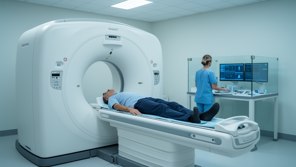
A shortened scanning range for prostate-specific membrane antigen PET/CT could save substantial scanner time, but a new retrospective study suggests the trade-off depends heavily on how far the scan is cut back and on each patient’s PSA level. The research, published in BMC Medical Imaging, examined what would be lost if a true whole-body scan were retrospectively trimmed into shorter, more conventional protocols.
The study, led by Falk Gühne and colleagues at a single center, included 200 patients with biochemically recurrent prostate cancer after initial curative therapy. All of them had undergone a 68Ga-PSMA PET/CT scan extending from the vertex of the skull to the toes, a true whole-body acquisition. The researchers then used retrospective subdivision into hypothetical partial-body measurement protocols to see which tumor manifestations would have gone undetected.
The rationale is practical. Most PET/CT scanners operate by measuring several bed positions or continuous motion with a limited axial field of view, so shortening the scan area saves time. The question was how much diagnostic information that time saving would cost.
Of the 200 patients, who had a median PSA of 1.1 ng/mL, 128 patients, or 64 percent, showed at least one structural correlate of biochemically recurrent prostate cancer in the true whole-body scan. The overall detection rate did not change for limited classic PET protocols or for chest/abdomen/pelvis measurements, but the picture changed for shorter protocols.
PSA emerged as a potentially useful guide for deciding how far the scan area could be restricted prospectively. Patients with PSA values of 0.75 ng/mL or below showed no diagnostic gain from whole-body imaging. In this cohort, that means a limitation to a chest/abdomen/pelvis scan would have been possible without losing information in 45 percent of patients.
The authors conclude that for a resource-saving use of routine PSMA PET/CT, a scan from the vertex to the mid-thigh appears sufficient for detecting relevant metastases. For patients with low PSA levels, a more focused protocol that includes only the chest/abdomen/pelvis region can be considered.
As a retrospective single-center analysis of 200 patients, the findings describe what would have been missed under hypothetical shortened protocols rather than outcomes from prospectively assigned scan ranges, and the authors note that PSA may contribute to prospective restriction of the scan area. Even so, the results offer a concrete framework for centers weighing scan time against diagnostic completeness in the follow-up of biochemically recurrent prostate cancer.
Subject of Research: The effect of reducing the scan extent of 68Ga-PSMA PET/CT on tumor detection in biochemically recurrent prostate cancer.
Article Title: Impact of reduced scan extent on tumor detection in 68Ga-PSMA PET/CT for biochemical recurrence of prostate cancer
Article References: Gühne, F., Küppers, A., Kühnel, C., Seifert, P., & Freesmeyer, M. (2026). Impact of reduced scan extent on tumor detection in 68Ga-PSMA PET/CT for biochemical recurrence of prostate cancer. BMC Medical Imaging, 26(1), Article 535. https://doi.org/10.1186/s12880-026-02940-w
Image Credits: AI Generated
DOI: 10.1186/s12880-026-02940-w
Keywords: PSMA PET/CT, prostate cancer, biochemical recurrence, whole-body imaging, scan protocols, PSA, tumor detection, field of view, chest abdomen pelvis, pelvic imaging, medical imaging, Impact
News Source: Nathaniel Bowman. (October 28, 2026). Shorter PET/CT scans may miss prostate cancer recurrence signs in some patients. Scienmag.