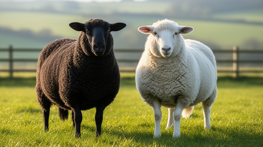
In the sweeping grasslands of Hungary’s Hortobágy National Park, one of Europe’s oldest sheep breeds wears its history in two striking colors. The Hortobágyi Racka, instantly recognizable by its long, spiraling V-shaped horns, comes in either a uniform jet black or a uniform chalk white coat. For centuries, shepherds have prized the breed for its remarkable hardiness, its ability to endure scorching summers and brutal winters alike. Now, a team of researchers has peered beneath the surface of those two coat colors, sequencing the genes active in the animals’ skin to ask a deceptively simple question: what, beyond pigment itself, actually differs between black and white Racka sheep? The answer, published in the journal Ecology and Evolution, turns out to reach far deeper than color, touching on ion channels, mitochondrial metabolism, and the cellular machinery of stress response.
The study, led by Putri Kusuma Astuti and colleagues at the University of Debrecen, focused on ten adult ewes of the Hortobágyi Racka breed, five black-coated and five white-coated, all of similar age and body weight, raised together under identical conditions and feeding management. Skin samples were collected from the posterior cervical region of each animal at an abattoir in northern Hungary in May 2023, preserved rapidly in RNAlater solution, and frozen at minus seventy degrees Celsius until analysis. By contrasting two coat colors within a single breed, the researchers deliberately minimized the confounding effects of breed-level genetic structure, allowing them to home in on expression differences genuinely associated with pigmentation rather than with broader ancestry.
The team used RNA sequencing, a technique that captures a snapshot of every gene being actively transcribed in a tissue at a given moment. Total RNA was extracted from the skin samples and converted into sequencing libraries using the QuantSeq 3′ mRNA-Seq kit, which focuses on the tail ends of messenger RNA molecules. The libraries were sequenced on an Illumina NovaSeq 6000 platform, generating more than fifty-four million reads across nine animals, as one black-coated sample failed to sequence. The reads were then aligned to the domestic sheep reference genome, the ARS-UI Ramb v3.0 assembly, using the STAR alignment tool, and gene-level counts were quantified with HTSeq. Sophisticated normalization and statistical modeling in the limma and edgeR frameworks, including the voom transformation that accounts for the peculiar mean-variance behavior of count data, allowed the researchers to identify genes whose expression differed significantly between the two color groups.
The results were strikingly one-sided. Of the 108 differentially expressed genes identified using a threshold of fold change greater than 1.5 and p less than 0.05, fully 83 were upregulated in the black-coated sheep, while only 25 were downregulated. The upregulated set was dominated by a tightly coordinated pigmentation network: melanogenesis enzymes such as TYR, TYRP1, and DCT; melanosome structural proteins including PMEL, MLANA, GPNMB, and TRPM1; melanosome transport genes like OCA2, SLC45A2, SLC24A5, and SLC24A4; and upstream melanocyte regulators including PAX3, IRF4, and RAB32. Nine of these genes, among them TYRP1, PMEL, TRPM1, MLANA, SLC24A4, SLC45A2, DCT, OCA2, and DDC, remained significant even after the most stringent multiple-testing correction. In other words, the skin of black Racka sheep is not merely darker; it is running an entire pigment-production program at far higher intensity.
Yet the story did not end with pigment. Functional enrichment analysis using Gene Ontology and KEGG pathway databases revealed that the differentially expressed genes clustered into pathways extending well beyond melanogenesis. The upregulated genes were significantly enriched in tyrosine metabolism, melanogenesis, metabolic pathways, thermogenesis, and oxidative phosphorylation, the latter being the mitochondrial process that generates most of a cell’s ATP. Gene-term networks and protein-protein interaction analysis built from the STRING database reinforced this picture, showing a coherent melanogenesis module centered on the TYR-DCT-MLANA axis, but also revealing separate modules devoted to mitochondrial and redox metabolism, involving genes such as NDUFS2, SLC25A4, QDPR, and AKR1B1, and to cell cycle and proteostasis signaling. Enriched molecular functions included calcium channel activity and calcium-potassium-sodium antiporter activity, reflecting the fact that melanin production inside melanosomes is tightly regulated by organellar ion homeostasis.
Why should coat color be entangled with cellular energy metabolism and stress biology? The authors point to a growing body of evidence that melanin synthesis is a biochemically demanding and redox-active process. Eumelanin, the pigment responsible for dark coloration, is a powerful antioxidant and an effective blocker of ultraviolet radiation, but producing it consumes cellular resources and can generate reactive oxygen species along the way. Pheomelanin, which produces lighter colors, is even more antioxidant-intensive, drawing on the cell’s stores of glutathione and cysteine. This means that the choice between dark and light pigmentation is not simply cosmetic; it carries real consequences for a skin cell’s oxidative balance, energy budget, and resilience under environmental stress such as intense sun and heat.
The study’s findings feed into an unresolved debate in livestock science about whether lighter or darker coats confer an advantage under a warming climate. Light-colored coats physically reflect more solar radiation and absorb less heat, which would seem advantageous in hot environments. Yet the evidence is mixed: some studies of grazing sheep in hot-arid conditions have found that black-coated animals maintained lower rectal temperatures than white-coated ones, while other research on goats under prolonged heat stress found white and mixed coats most adaptable. A recent gene-editing experiment in Holstein cattle, in which the PMEL gene was modified to dilute coat color, showed that lighter calves absorbed significantly less heat and light than their darker counterparts. The Racka data do not settle the question, but they suggest a mechanism by which dark pigmentation could carry its own compensatory benefits: the upregulation of genes like TRPM1, implicated in DNA repair and calcium signaling during heat stress, and TYRP1, reported to regulate oxidative stress and support cellular adaptation in extreme conditions.
There is also an intriguing immunological dimension. Melanocytes, the pigment-producing cells of the skin, are increasingly understood as active participants in immune signaling. They can present antigens to T lymphocytes, express immune-interaction molecules such as ICAM-1 and CD40, and release cytokines that shape inflammatory responses. Highly melanized melanocytes may suppress inflammation more effectively, partly through the immunosuppressive effects of L-DOPA and related compounds, whereas lightly pigmented skin may mount stronger cytokine-mediated immune signaling. The researchers also note that pigment regulation and the body’s stress-hormone system share biochemical machinery, notably the proopiomelanocortin molecule, which feeds into both pigmentation and cortisol synthesis. This overlap raises the possibility that coat color and stress physiology are coupled through shared hormonal pathways, a hypothesis the authors frame as speculative but testable.
The team validated their sequencing results using quantitative reverse-transcription PCR on ten selected genes spanning pigmentation, immune, and metabolic functions. Eight of the ten genes were confirmed to be significantly more highly expressed in black sheep skin, with TYRP1 showing the largest difference between the groups. The authors are careful, however, to spell out the limitations of their work. With only four black and five white animals, statistical power is modest, and the study included no direct measurements of heat tolerance, UV response, oxidative stress markers, or skin temperature. The links between pigmentation and adaptation remain, in their words, hypothesis-generating rather than causal, awaiting protein-level assays and controlled heat-challenge experiments in larger populations.
Even with those caveats, the study delivers a compelling molecular portrait of a famous breed and a provocative idea: that the color of a sheep’s fleece is the visible tip of a much larger biological iceberg. Beneath the black and white coats of the Hortobágyi Racka lie coordinated differences in ion transport, mitochondrial bioenergetics, redox buffering, and proteostasis, suggesting that pigmentation and environmental resilience may have evolved together. As climate change intensifies heat stress on livestock worldwide, understanding how coat color genes intertwine with cellular stress machinery could help breeders select animals not just for appearance, but for the hidden physiology that keeps them thriving in an increasingly hostile environment.
Subject of Research: Transcriptomic differences between black- and white-coated Hortobágyi Racka sheep skin
Article Title: Coat Color‐Associated Transcriptomic Differences of Black and White Hortobágyi Racka Sheep Using the RNA‐Seq Technique
Article References: Astuti, P. K., Bagi, Z., Bodrogi, L., & Kusza, S. (2026). Coat Color‐Associated Transcriptomic Differences of Black and White Hortobágyi Racka Sheep Using the RNA‐Seq Technique. Ecology and Evolution, 16(10), Article e74397. https://doi.org/10.1002/ece3.74397
Image Credits: AI Generated
DOI: 10.1002/ece3.74397
Keywords: Hortobágyi Racka sheep, coat color, RNA-seq, melanogenesis, transcriptomics, pigmentation, heat stress, oxidative stress, mitochondrial metabolism, TYRP1, melanocytes, livestock adaptation
News Source: Juliet Wilcox. (October 9, 2026). Sheep Skin Genes Reveal Hidden Biology Behind Black and White Coats. Scienmag.