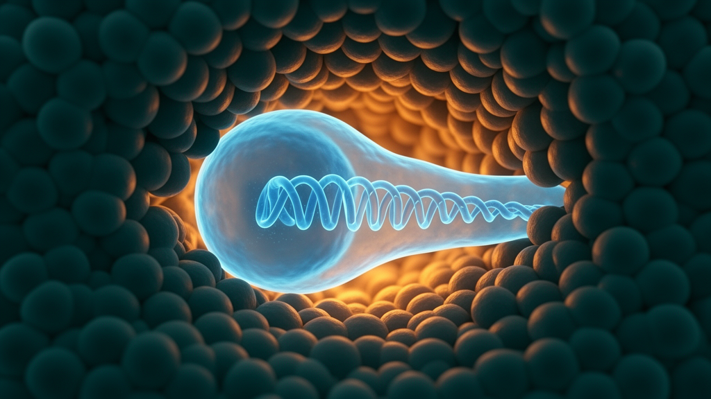
Every developing embryo is a construction site in which cells must travel long distances through spaces that are often far too small for them. As a migrating cell pushes through a narrow gap, its nucleus — typically the stiffest and most fragile organelle in the cell — is forced to deform dramatically. In laboratory dishes, this kind of squeezing has been shown to rupture the nuclear envelope, the double membrane that shields the genome, and to trigger DNA damage that can push cells toward cancerous behaviour. Whether the same hazards apply inside a living, normally developing organism has been far less clear. A new study published in Nature Cell Biology by Hanna-Maria Häkkinen, Elena Scarpa and colleagues at the University of Cambridge and their collaborators now shows that at least one remarkable embryonic cell type has evolved a way to squeeze through extremely tight tissue corridors without breaking its genome.
The team turned to the zebrafish neural crest, a population of pluripotent stem-like cells that arises along the dorsal side of the neural tube and migrates extensively throughout the embryo before differentiating into an astonishing variety of tissues, including pigment cells, neurons and craniofacial structures. Crucially, neural crest cells encounter very different physical environments depending on where they migrate along the head-to-tail axis. Cranial neural crest cells travel through loosely organised, fluid-filled spaces behind the eye, whereas trunk neural crest cells must invade narrow inter-tissue corridors between the spinal cord and the somites, the segmented blocks of tissue that will form muscle. This natural gradient of confinement provided the researchers with a built-in experiment: cells migrating through the same embryo under different degrees of physical constraint.
Using live imaging of embryos carrying fluorescent reporters that label neural crest nuclei and membranes, together with injected fluorescent dextran that outlines the extracellular spaces, the researchers quantified nuclear shapes as cells migrated. They found that cranial and anterior trunk neural crest nuclei remained largely circular and isotropic throughout migration. In contrast, mid-trunk and posterior trunk neural crest cells underwent repeated, dramatic nuclear deformations, with circularity dropping sharply as each cell squeezed beneath the somite. The severity and duration of these deformation events increased progressively along the anterior-to-posterior axis of the embryo, while cell shape itself changed little — a strong indication that the nucleus, not the cytoplasm, was bearing the brunt of the mechanical stress.
To confirm that tissue confinement was truly the cause of these nuclear shape changes, the team perturbed the surrounding tissues in two independent ways. First, they used the spadetail mutant, in which somite formation is defective and the spaces along the trunk migratory path are significantly wider. In these embryos, trunk neural crest nuclei no longer deformed as they entered the migratory zone. Second, they used femtosecond pulsed infrared laser ablation to physically remove a portion of the presomitic mesoderm, creating a local gap in the somite organisation while leaving the neural tube, notochord and premigratory neural crest intact. Again, cells migrating through the ablated region showed markedly less nuclear deformation than cells passing through adjacent, untouched somites. Together, these orthogonal genetic and mechanical manipulations established that confinement imposed by the somite tissue is what drives the striking nuclear distortions.
The next question was whether these deformations compromise nuclear integrity. In cultured cells, migration through pores smaller than a critical threshold of roughly three micrometres causes transient ruptures of the nuclear envelope, exposing chromatin to the cytoplasm and allowing DNA repair factors to leak away. The researchers imaged trunk neural crest cells expressing a nuclear-localised emerald GFP and observed a small, transient leakage of the reporter from the nucleus into the cytoplasm at the moment of maximum deformation — a decrease of only about five per cent in nuclear intensity. Crucially, however, two independent nuclear envelope rupture reporters, BAF-mCherry and cGAS-EGFP, showed no accumulation at the nuclear periphery of deforming cells, even though both reporters readily flagged experimentally induced ruptures. Scanning electron microscopy of trunk cross-sections confirmed that the nuclear envelope remained intact in migrating cells, and revealed a wide distribution of nuclear pore widths, suggesting that the leakage may occur through mechanically stretched nuclear pores rather than through catastrophic envelope failure.
Even more striking was what the team found when they looked for DNA damage. Immunostaining for phosphorylated γH2AX, a canonical marker of DNA double-strand breaks, revealed that migrating trunk neural crest cells did not show increased damage compared with premigratory cells — and in the most strongly deformed posterior population, γH2AX levels were actually significantly lower. A live reporter based on the DNA damage protein 53BP1, expressed from injected mRNA, allowed the researchers to track damage foci in real time. Foci, when they appeared, resolved within minutes, and the intensity of the 53BP1 signal remained constant for up to an hour before and after each maximum deformation event. Correlation analysis found no relationship between how strongly a nucleus was squeezed and how much DNA damage response activity it displayed.
To test whether the softness of embryonic tissue was the reason for this protection, the researchers measured the stiffness of live zebrafish trunk tissue using atomic force microscopy and found it to be remarkably soft, at roughly 0.4 kilopascals — orders of magnitude softer than the stiff polydimethylsiloxane devices typically used in microfabrication studies. They then cultured primary trunk neural crest cells in PDMS pillar forests with three-micrometre gaps, imposing far harsher, non-deformable confinement than the embryo provides. Even under these rigid conditions, the cells deformed their nuclei extensively yet still showed no increase in 53BP1 signal, no correlation between deformation and damage, and no accumulation of double-strand breaks. The protection, in other words, is intrinsic to the cells rather than a gift of their soft surroundings.
The search for the mechanism behind this resilience led to the nuclear lamina, the protein meshwork that underlies the nuclear envelope and largely determines nuclear stiffness. Early zebrafish embryos express little or no LaminA/C, and the team found that LaminB2 was the most enriched lamin in trunk neural crest cells. Remarkably, confined migratory trunk cells significantly reduced LaminB2 at the nuclear envelope, while non-confined cranial and anterior trunk cells did not. Functional perturbations revealed that LaminB2 acts as a regulator of nuclear deformability: knocking it down slightly shortened the duration of deformation events, whereas sustained overexpression of a Halo-tagged LaminB2 caused nuclei to remain persistently distorted and was associated with a mild increase in overall 53BP1 intensity. Fine-tuning LaminB2 levels, the authors conclude, allows the nucleus to soften and recover efficiently as it passes through constrictions.
The final piece of the puzzle came from transcriptomics. Using a photoconvertible H2B-Dendra2 line, the researchers labelled mid-trunk neural crest nuclei either before migration or at the endpoint of confined migration, sorted the cells and performed low-input bulk RNA sequencing. The single most upregulated biological process in cells that had completed confined migration was the DNA damage response, encompassing around seventy genes drawn from homologous recombination, non-homologous end joining and checkpoint signalling pathways. This suggests that these embryonic stem-like cells are intrinsically armed with a comprehensive repair programme, primed to deal with any lesions that might arise, even though measurable damage remains low. The authors note that inhibiting BMP signalling, which has been linked to DNA damage protection in other zebrafish contexts, did not affect 53BP1 accumulation, leaving open the question of how cells sense confinement to activate the programme.
The findings carry implications well beyond developmental biology. Neural crest-derived cancers, including melanoma and neuroblastoma, often reactivate embryonic migration programmes, and neuroblastoma predisposition genes cluster in DNA repair and checkpoint pathways, with growing evidence that these tumours originate in utero during early trunk neural crest development. Understanding how multipotent embryonic cells protect their genomes while navigating physically hostile tissue landscapes may therefore illuminate both the robustness of normal development and the mechanical origins of genomic instability in cancer. For now, the zebrafish neural crest stands as an elegant demonstration that evolution has equipped migrating embryonic cells with a layered defence — a softened, adaptable nucleus, an envelope that bends rather than breaks, and a repair toolkit switched on in anticipation of stress.
Subject of Research: DNA damage protection in confined in vivo migration of zebrafish neural crest cells
Article Title: In vivo DNA damage protection during cell migration across confining embryonic tissue environments
Article References: Häkkinen, H.-M., Villaseca, S., Alhashem, Z., Hamidzadeh, A., Chomiczewski, S., Desevedavy, M., Leleux, S., Liu, Y.-H., Becker, J. M., Htun, M. R., Gallo, F., El-Zohiry, D., Petre, V., Stefanowski, K., Franze, K., Renkawitz, J., & Scarpa, E. (2026). In vivo DNA damage protection during cell migration across confining embryonic tissue environments. Nature Cell Biology. https://doi.org/10.1038/s41556-026-02065-w
Image Credits: AI Generated
DOI: 10.1038/s41556-026-02065-w
Keywords: zebrafish, neural crest, cell migration, nuclear deformation, nuclear envelope, DNA damage, LaminB2, confinement, embryonic development, 53BP1, RNA-seq, nuclear lamina
News Source: Drew Townsend. (October 9, 2026). Embryonic Cells Squeeze Through Tight Tissues Without Breaking Their DNA. Scienmag.