
In the rapidly evolving field of medical imaging, the incorporation of artificial intelligence (AI) is reshaping how clinicians assess and interpret images, particularly in the context of hepatobiliary diseases. Recently published in the esteemed journal, Oncotarget, an editorial titled “Deep learning-based uncertainty quantification for quality assurance in hepatobiliary imaging-based techniques” sheds light on significant advancements in this sector. The editorial, authored by Dr. Yashbir Singh and his colleagues from Mayo Clinic, explores the critical role of uncertainty quantification in enhancing the detection of liver pathologies, which can often be complex and challenging to diagnose.
AI’s potential lies not only in its capacity to process images with speed and accuracy, but also in its ability to self-assess confidence in its diagnostic suggestions. This innovative concept of uncertainty quantification empowers AI systems to highlight scans where uncertainty is high. For instance, when AI algorithms process liver scans, they examine various features and patterns, subsequently generating confidence scores that indicate how certain they are about their findings. This added layer of interpretive clarity is particularly vital in high-stakes clinical settings, where the identification of conditions like liver cancer can hinge on nuanced details within imaging results.
Liver imaging has historically presented numerous challenges due to the organ’s intricate anatomical structures and the variability in image quality. Factors such as patient anatomy, the presence of liver damage, and technical aspects of imaging technology can obscure the visibility of small tumors. In response to these challenges, modern AI models, including those discussed in the editorial, utilize advanced deep learning techniques to effectively analyze imaging data while providing concurrent uncertainty metrics. This dual functionality enhances clinical decision-making, ensuring that physicians can make more informed evaluations when interpreting results.
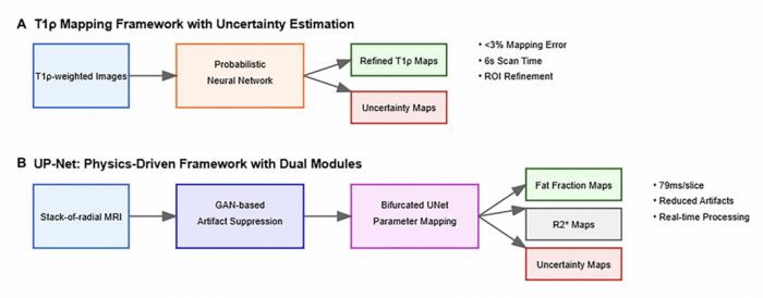
One notable model highlighted in the editorial is the Anisotropic Hybrid Network, or AHUNet, which adeptly handles both two-dimensional and three-dimensional liver scans. The strength of AHUNet lies in its ability to identify specific areas within an image where the algorithm is confident versus where it harbors uncertainty. By utilizing such models, clinicians can direct their focus toward scans that require additional scrutiny, significantly lowering the risk of misdiagnosis, particularly among patients with underlying liver diseases.
The editorial also outlines the transformative potential of AI tools in the context of liver imaging through the use of frameworks that can automatically analyze and quantify liver fat. This capability not only enhances diagnostic accuracy but also allows for rapid assessments, which are crucial in busy clinical environments. For instance, some AI systems can examine ultrasound images and provide both a diagnostic output and a corresponding confidence rating within a fraction of the time it would take a human radiologist. This speed and efficiency not only alleviate the workload on radiologists but also promote better overall patient care.
Moreover, the implications of these advancements extend beyond urban centers to smaller clinics, where access to specialized hepatobiliary expertise may be limited. AI’s ability to flag uncertain findings can ensure that questionable results are promptly escalated to larger medical institutions for further evaluation. Such a system not only enhances diagnostic capabilities but also democratizes access to quality healthcare, enabling even rural and underserved populations to benefit from advancements in medical imaging technology.
As these tools gain traction, there is a pressing need for standardization in radiological reporting methods. The authors of the editorial advocate for the development of standardized reporting templates that incorporate uncertainty metrics side-by-side with conventional imaging findings. This integration is imperative for cultivating a culture where interpretative confidence is communicated clearly, fostering a scenario where clinicians and patients can make collaborative, informed decisions about treatment pathways.
The potential impact of AI in radiology cannot be overstated. As AI tools become adept at recognizing when they should variably adjust their confidence levels, they offer clinicians a robust mechanism for enhancing accuracy in liver cancer detection and the monitoring of liver diseases. The article posits that uncertainty-aware AI may soon evolve into a cornerstone of conventional medical imaging practices, underpinning swift and precise decision-making processes in liver disease management.
Continuing advancements in deep learning technology promise to enhance diagnostic workflows, enabling not only quicker turnaround times for results but also improved accuracy that could ultimately save lives. The authors emphasize the importance of ongoing collaboration between AI developers and healthcare professionals to ensure that these tools are effectively integrated into everyday medical practice, maximizing their utility and effectiveness.
In summary, the integration of deep learning and uncertainty quantification within hepatobiliary imaging signifies a monumental leap forward in medical diagnostics. The synergy between human expertise and AI-driven analysis offers an unprecedented opportunity to enhance clinical outcomes, streamline workflows, and ultimately revolutionize patient care in hepatobiliary medicine. As this technology matures, it is poised to redefine the standards and practices associated with liver disease detection, leading to better prognosis and treatment options for patients.
Furthermore, as the scientific community eagerly anticipates the application of these technologies in routine practice, it remains crucial to address ethical considerations surrounding AI in healthcare. Transparency in AI decision-making processes can foster trust among users and patients alike, ensuring that AI’s integration serves the overarching goal of improving health outcomes while respecting patient autonomy and privacy.
The future of hepatobiliary imaging is set to be characterized by new dimensions of reliability and efficiency, ensuring that even the most subtle clinical findings do not evade detection, ultimately reshaping the landscape of cancer diagnostics in significant and profoundly positive ways.
Subject of Research: Not applicable
Article Title: Deep learning-based uncertainty quantification for quality assurance in hepatobiliary imaging-based techniques
News Publication Date: April 4, 2025
Web References: Not available
References: Not available
Image Credits: Copyright: © 2025 Singh et al.
Keywords: cancer, deep learning, uncertainty quantification, radiology, hepatobiliary imaging
Tags: AI in liver pathologyconfidence scoring in diagnosticsdeep learning in healthcarehepatobiliary diseases diagnosishigh-stakes clinical decision-makinginterpretive clarity in medical imagingliver cancer detectionMayo Clinic researchmedical imaging advancementsquality assurance in imaging techniquesself-assessing AIuncertainty quantification in AI

