


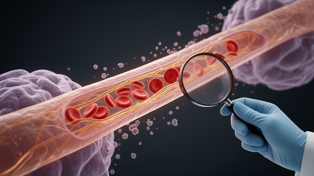
Alzheimer’s disease research has long concentrated on the two parenchymal hallmarks of the condition—amyloid-β plaques and tau tangles—as the principal drivers of neurodegeneration. But a new Perspective article argues that this focus has left a critical blind spot: the brain’s blood vessels. Writing in PLOS Biology, Thomas S. Lewis, Jessica Wagner, and Jonas J. Neher of LMU Munich and the German Center for Neurodegenerative Diseases contend that vascular dysfunction is emerging as a significant and independent contributor to neuronal damage, yet the field’s powerful molecular profiling tools have raced far ahead of its ability to say what those molecular changes actually do to vessel function.
The core problem, the authors argue, is one of inference. Molecular profiling studies have mapped extensive alterations in the cerebral blood vessels of Alzheimer’s patients and animal models, cataloguing changes in gene expression, extracellular matrix composition, and cellular behavior. What these studies have not done, they stress, is proven a causal role for these alterations in pathology. Molecular or cellular abnormalities alone cannot establish whether, or to what extent, a vessel is functionally impaired. In their view, vascular dysfunction is frequently inferred from histological or molecular phenotypes rather than quantified physiologically—a limitation they describe as critical for the field.
The case for taking vascular dysfunction seriously rests on timing. The authors note that neurovascular dysfunction is among the earliest detectable abnormalities in patients with Alzheimer’s disease and may precede the biomarker changes that reflect amyloid-β and tau pathology. Reductions in cerebral blood flow and impaired neurovascular coupling—the mechanism by which local blood supply is matched to neuronal activity—are already present at prodromal stages of the disease. By advanced stages, regional cerebral blood flow may decrease by up to 45 percent in patients. These observations, the authors write, justify treating vascular dysfunction as a potential contributor to disease progression rather than as a secondary consequence of neuronal pathology.
Yet timing alone does not establish mechanism. The authors acknowledge that these observations do not identify what actually impairs vascular function. Despite recent advances in molecular and cellular characterization of cerebral blood vessels, little is known about the drivers of these alterations, how closely they are linked to protein aggregation, how they progress over the course of disease, or how they translate into measurable changes in vascular function. Distinguishing whether vascular dysfunction causes, amplifies, or merely follows other hallmarks of Alzheimer’s disease is, they argue, essential for developing earlier diagnostic biomarkers and for prioritizing vascular changes as therapeutic targets rather than mere correlates of the disease.
One of the most detailed examples the authors discuss is the vascular extracellular matrix, the fibrous scaffold surrounding blood vessels. Extensive remodeling of this matrix has been reported in both aging and Alzheimer’s disease, involving alterations in collagens, laminins, fibronectin, proteoglycans, and basement membrane proteins. Many of these proteins can be detected in cerebrospinal fluid, raising the possibility that they could serve as biomarkers of vascular pathology. The matrix is far more than passive scaffolding: it regulates endothelial signaling, vascular smooth muscle function, basement membrane integrity, arterial compliance, and perivascular clearance, so changes in its composition may have direct consequences for how vessels behave.
The authors sketch a plausible mechanistic chain. Changes in collagen organization, elastin integrity, and basement membrane composition may increase arterial stiffness. Meanwhile, amyloid-β deposition within vessel walls may impair vessel elasticity, disrupt the clearance of waste products along perivascular pathways, and initiate inflammatory cascades. Together, they write, these changes provide a plausible explanation for the altered neurovascular function and hypoperfusion observed in patients with Alzheimer’s disease. But they issue a sharp caveat: biological plausibility should not be mistaken for demonstrated physiological dysfunction. The functional consequences of these molecular and structural changes have rarely been measured directly.
Closing that gap, the authors argue, will require measuring vascular properties across different spatial and temporal scales—and no single imaging modality can do the whole job. Two-photon microscopy enables real-time visualization of individual arterioles, capillaries, and vascular cells, revealing the mechanisms of neurovascular coupling, vasoconstriction, and vasodilation, but it interrogates only small cortical areas and therefore offers a highly localized view. Functional MRI can visualize blood flow throughout the entire brain, but at substantially lower spatial resolution. Notably, neither approach can determine changes in the passive properties of vessels, such as pulse-wave velocity, a measure of vascular stiffness.
That measurement gap is where the authors highlight high-frequency ultrasound as a complementary technology. By acquiring thousands of frames per second, high-frequency ultrasound can resolve individual cardiac cycles, enabling estimates of arterial stiffness and vascular resistance. A contrast-enhanced variant extends the assessment to microvascular perfusion and to computational reconstruction of the functional cerebral vascular network, from meningeal vessels down to the microvasculature. In the authors’ framing, this places ultrasound at a scale between cellular imaging by two-photon microscopy and whole-brain measurements by MRI. They caution, however, that comprehensive analysis will require integration across modalities, which is technically demanding: it demands complementary equipment and expertise, as well as anatomical co-registration of datasets from the same animal, often across different facilities.
Because establishing temporal order and causality is central to their argument, the authors see a key role for animal models. Although genetic mouse models do not recapitulate the full complexity of human Alzheimer’s disease, they permit vascular alterations to be monitored and manipulated longitudinally, and they reproduce age-associated changes that the authors consider of major importance for understanding sporadic disease. These models reproduce selected components of Alzheimer’s pathology, particularly amyloid-β deposition and cerebral amyloid angiopathy, and some also exhibit impaired neurovascular coupling and reduced cerebral blood flow. Defining mechanistic links between molecular remodeling and vascular dysfunction in such models, they argue, can then guide targeted studies in patients.
The authors’ proposed way forward is a longitudinal research program that links molecular and structural vascular changes to vascular mechanics, hemodynamics, tissue perfusion, and oxygen delivery. Cellular responses, vascular biomechanics, network hemodynamics, tissue perfusion, and oxygenation capture distinct but interconnected aspects of vascular function, so combining measurements across these scales is essential. Modern imaging techniques, complemented by histological and spatial omics approaches, could provide a comprehensive map of where and how blood vessels are affected, how those changes alter vascular network function, and how they affect brain homeostasis. Such an approach, they suggest, could identify earlier disease biomarkers, strengthen mechanistic understanding of vascular contributions to neurodegeneration, reveal therapeutic targets, and enable spatial and temporal monitoring of interventions—including identifying the disease stages at which interventions to preserve or restore adequate cerebral perfusion could be tested. Although their focus is Alzheimer’s disease, they note the framework may also apply to other neurodegenerative diseases and to healthy aging. Their concluding message is blunt: understanding what changes occur within the brain’s vessels is no longer sufficient; researchers must determine how those changes affect vascular function and brain perfusion, and which components can be targeted to preserve or restore brain health.
Subject of Research: Vascular dysfunction and molecular profiling of cerebral blood vessels in Alzheimer's disease research
Article Title: Alzheimer’s disease research must connect vascular molecular phenotypes with physiology
Article References: Lewis, T. S., Wagner, J., & Neher, J. J. (2026). Alzheimer’s disease research must connect vascular molecular phenotypes with physiology. PLOS Biology, 24(10), e3004051. https://doi.org/10.1371/journal.pbio.3004051
Image Credits: AI Generated
DOI: 10.1371/journal.pbio.3004051
Keywords: Alzheimer's disease, cerebral blood vessels, vascular dysfunction, neurovascular coupling, cerebral blood flow, extracellular matrix, arterial stiffness, high-frequency ultrasound, two-photon microscopy, functional MRI, amyloid-β, PLOS Biology
News Source: Cassandra Pierce. (October 28, 2026). Scientists Say Alzheimer’s Vessel Findings Lack Proof of Function. Scienmag.



