New approach helps researchers determine the resolution of cryo-EM, leading to better methods of imaging proteins
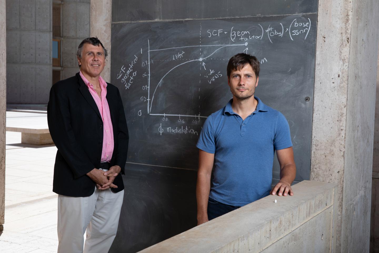
Credit: Salk Institute
In 2017, Salk scientists reported that tilting a frozen protein sample as it sat under an electron microscope was an effective approach to acquiring better information about its structure and helping researchers understand a host of diseases ranging from HIV to cancer. Now, they have developed a mathematical framework that underlies some of those initial observations.
Their new study, published in Progress in Biophysics and Molecular Biology on September 13, 2019, provides a foundation for quantitatively determining how differences in viewing angles affect the resulting 3D structures of proteins, and could help other researchers determine the best setup for experiments to improve the imaging technique called cryo-EM.
“This provides a quantitative understanding for why variations in viewing angles affect the quality of resulting 3D structures of proteins, and where we could do better to improve the data,” says Dmitry Lyumkis, a Salk assistant professor of genetics and coauthor of the new work. “These kinds of theoretical frameworks are important to understanding precisely how information is attenuated due to imperfections associated with the imaging experiment, which will lead us to eventually get better structures out of cryo-EM data.”
In cryo-EM, or cryogenic electron microscopy, proteins are rapidly frozen in their natural form before being bombarded with electrons. By detecting how the electrons scatter when they hit the sample, researchers can determine the molecular structure of the protein or protein complex. Compared to other imaging methods, it’s easier for scientists to prepare proteins for cryo-EM, and the technique can potentially address a broader set of questions in structural biology. However, a long-standing problem in cryo-EM is that proteins tend to stick to the top or bottom of the sample grid that they’re prepared on. These select orientations mean that researchers can’t always see a protein’s structure from every angle. Tilting the sample, Lyumkis and his colleagues found in 2017, helped solve this problem.
“We knew that qualitatively, tilting improved the data in some cases,” says Lyumkis. “What we didn’t know was exactly the extent to which the structures can be affected by variations in the viewing angle.”
Recently, Philip Baldwin, a senior staff researcher at Salk and the paper’s coauthor, was examining a set of cryo-EM data collected at different viewing angles when he noticed that such variations affected the overall resolution of the resulting protein structure. After some calculations, he realized that the association between the viewing angle and resolution was generalizable to all cryo-EM experiments.
The new formula lets researchers calculate, for any protein at any tilt angle, a number called the sampling compensation factor, or SCF. The closer the SCF value is to 1, the more complete the protein’s structure. If the SCF is 0.5 instead of 1, either the data is incomplete, or researchers must collect data for twice as long to get the same structural resolution. By calculating SCF values ahead of an experiment, scientists can optimize their tilt angle and data collection time.
The new quantitative formulations also helped Lyumkis and Baldwin compute just how incomplete some cryo-EM datasets are. Previously, they might have had to eyeball a set of data and guess whether it was a good or bad approximation of a protein’s structure. Now, the SCF can tell them that numerically.
“It’s very handy,” says Baldwin. “Basically, this formula tells you if you have very bad regions of the protein from which you didn’t collect data.”
Lyumkis and Baldwin hope that using the formula to assess cryo-EM results–which involves a simple calculation or piece of code–will become standard and help guide experiments and new approaches to cryo-EM. This would lead to faster discoveries in basic biological sciences and in drug development.
The work reflects a growing trend at both the Salk Institute and elsewhere toward integrating computational approaches into biology research.
###
Lyumkis and Baldwin were supported by grants from the National Institutes of Health.
About the Salk Institute for Biological Studies:
Every cure has a starting point. The Salk Institute embodies Jonas Salk’s mission to dare to make dreams into reality. Its internationally renowned and award-winning scientists explore the very foundations of life, seeking new understandings in neuroscience, genetics, immunology, plant biology and more. The Institute is an independent nonprofit organization and architectural landmark: small by choice, intimate by nature and fearless in the face of any challenge. Be it cancer or Alzheimer’s, aging or diabetes, Salk is where cures begin. Learn more at: salk.edu.
Media Contact
Salk Communications
[email protected]
Original Source
https:/
Related Journal Article
http://dx.