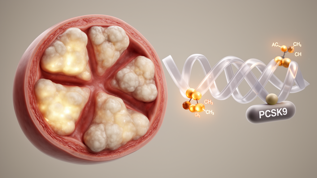
Calcific aortic valve disease has long been one of cardiology’s most stubborn problems: a slowly stiffening aortic valve that narrows the outflow of the heart, with no drug able to stop it. Once symptoms appear, the only effective remedy remains surgical or transcatheter valve replacement, procedures that are palliative rather than curative. Even lipid-lowering therapies, which transformed the treatment of atherosclerotic heart disease, have failed to halt valve calcification in clinical trials. Now a study published in iScience offers a fresh molecular explanation for why the valve turns to bone, and it points to an unexpected target: an enzyme that chemically decorates messenger RNA.
The research, led by Yinghuan Liu, Xianying Li, Tong Zhou, Jianhua Xie and colleagues under the supervision of Yongjun Wang, Dong Han and Lifu Li, focuses on N-acetyltransferase 10, or NAT10, the only known mammalian enzyme that installs N4-acetylcytidine, abbreviated ac4C, onto cytidine residues within mRNA. This chemical acetylation is a member of the expanding family of epitranscriptomic marks, modifications that do not change the genetic sequence itself but alter how RNA molecules behave, influencing their stability, localization, translation efficiency and fidelity. By stabilizing particular transcripts, ac4C can effectively turn up the volume of specific genes without the cell ever touching its DNA.
The team began by asking whether NAT10 is present in diseased valves. Analyzing tissue from 50 patients with calcific aortic valves and 50 non-calcified controls, they found that NAT10 mRNA and protein were both significantly elevated in the diseased tissue. A parallel increase appeared in a mouse model, in which apolipoprotein E-deficient mice fed a high-cholesterol diet for 24 weeks develop aortic valve calcification that mirrors the human condition. Dot blot assays measuring total RNA acetylation revealed that global ac4C levels rose alongside NAT10 in both human and mouse calcified valves, suggesting that the enzyme’s catalytic output, not merely its abundance, tracks with disease.
Cellular studies sharpened the picture. The chief cellular culprit in valve calcification is the human valvular interstitial cell, or hVIC, which under pathological stress abandons its usual quiescent state and transforms into an osteoblast-like cell that lays down mineral. When the researchers isolated hVICs and valve endothelial cells from patients, NAT10 was substantially higher in the interstitial cells, identifying them as the primary source of the enzyme in diseased valves. When hVICs were bathed in osteogenic medium, NAT10 expression climbed in a time-dependent manner together with the osteogenic marker osterix. Silencing NAT10 blunted the appearance of osteogenic markers, calcified nodule formation and calcium deposition, while forcing NAT10 overexpression intensified all of these features, effects that held true in cells from both diseased and healthy donors.
Correlation analyses in a second, independent validation cohort of 50 patients linked NAT10 to disease severity in a clinically meaningful way. NAT10 expression rose in step with the osteogenic markers osterix and osteopontin, and higher NAT10 levels were associated with a smaller aortic valve area and greater mean and peak transvalvular pressure gradients on echocardiography. In other words, the more NAT10 a patient’s valve expressed, the worse the valve functioned, positioning the enzyme as both a potential biomarker and a therapeutic target.
To find what NAT10 was actually doing downstream, the investigators performed a rigorous three-way screen. They intersected genes downregulated when NAT10 was knocked down in hVICs, established NAT10 perturbation targets from the RM2Target database, and aortic valve calcification genes curated in the Open Targets Platform. The overlap narrowed to just seven candidates: COL1A1, PCSK9, SCG2, UBE2C, FN1, THBS2 and SERPINE1. Rescue experiments then delivered a decisive verdict. Knocking down PCSK9, but none of the other six genes, almost completely abolished the pro-calcific phenotype driven by NAT10 overexpression, establishing PCSK9 as the dominant functional effector of the pathway.
PCSK9 is best known as a cholesterol regulator, famous for degrading low-density lipoprotein receptors in the liver and for the blockbuster antibody drugs that block it. But recent genetic and experimental work has implicated PCSK9 directly in valve calcification as well. The new study shows that NAT10 regulates PCSK9 post-transcriptionally. RNA decay assays using actinomycin D to halt transcription demonstrated that NAT10 knockdown shortened the half-life of PCSK9 mRNA, while NAT10 overexpression prolonged it, with corresponding changes in PCSK9 protein production and secretion. In human calcific valves, NAT10 and PCSK9 expression correlated positively, tying the molecular mechanism to clinical tissue.
The mechanistic proof that ac4C is the responsible mark came from a series of elegant experiments. RNA immunoprecipitation showed that PCSK9 mRNA is bound by both NAT10 and anti-ac4C antibodies, and that NAT10 depletion reduces the ac4C signal on the transcript. A predicted acetylation motif within positions 1886 to 1900 of the PCSK9 coding sequence was tested with luciferase reporters: mutating the cytosines in that motif to thymines made the reporter insensitive to NAT10 knockdown, whereas the wild-type reporter responded strongly. The researchers also deployed a catalytically dead NAT10 mutant, G641E, which retains expression but cannot acetylate RNA. Only the wild-type enzyme, not the mutant, boosted PCSK9 stability and production, proving that the acetyltransferase activity itself is essential. Control experiments confirmed that knockdown of NAT10 did not perturb other components of the ac4C machinery, including the cofactor THUMPD1, the reader NOP58 and the putative eraser SIRT7, underscoring the specificity of the effect.
The most clinically compelling results came from the mouse experiments. Apolipoprotein E-deficient mice on a high-cholesterol diet were treated either with remodelin, a small-molecule NAT10 inhibitor, or with NP1192, a first-in-class PROTAC degrader that selectively drags NAT10 into the cellular destruction machinery. Because remodelin is known to interact with multiple cellular targets, the PROTAC served as an orthogonal check on specificity. Both compounds partially reversed the echocardiographic hallmarks of aortic stenosis, reduced valve leaflet thickness and calcium deposition on histology, suppressed the osteogenic markers osterix and osteopontin, and lowered NAT10 protein and global ac4C levels in valve tissue. Crucially, blood glucose, total cholesterol, LDL, triglycerides and circulating PCSK9 were unchanged by treatment, indicating that the benefit arose from local action within the valve rather than from systemic lipid lowering, and that the anti-calcific effect was on-target rather than a metabolic side effect.
The study is not without caveats, which the authors acknowledge candidly. Calcific valve disease involves endothelial cells and macrophages in addition to interstitial cells, and NAT10’s role in those compartments was not explored; cell-type-specific knockout models will be needed to disentangle the contributions. Transcriptome-wide mapping of ac4C targets was not performed, so other NAT10-regulated transcripts may contribute to the phenotype. The upstream signals that raise NAT10 expression in the diseased valve, whether mechanical stress, lipid retention, oxidative stress or inflammatory cytokines, remain unidentified. Even so, the work delivers something the field has lacked: a concrete, druggable epitranscriptomic axis, NAT10-ac4C-PCSK9, that connects RNA chemistry to the bone-like transformation of the aortic valve. If future clinical studies confirm that NAT10 degraders such as NP1192 can slow or stop valve calcification in patients, the humble acetyl group on a single cytidine may turn out to be the handle medicine has been searching for on one of cardiology’s hardest diseases.
Subject of Research: The role of NAT10-mediated mRNA N4-acetylcytidine modification of PCSK9 in calcific aortic valve disease
Article Title: NAT10 exacerbates aortic valve calcification via PCSK9 mRNA ac4C acetylation
Article References: Liu, Y., Li, X., Zhou, T., Xie, J., Jiang, H., Zheng, B., Peng, J., Li, L., Han, D., & Wang, Y. (2026). NAT10 exacerbates aortic valve calcification via PCSK9 mRNA ac4C acetylation. iScience, 29(11), Article 117812. https://doi.org/10.1016/j.isci.2026.117812
Image Credits: AI Generated
DOI: 10.1016/j.isci.2026.117812
Keywords: NAT10, ac4C, PCSK9, aortic valve calcification, calcific aortic valve disease, epitranscriptomics, valvular interstitial cells, RNA modification, PROTAC, NP1192, osteogenic differentiation, mRNA stability
News Source: Denise Maddox. (October 14, 2026). RNA acetylation enzyme NAT10 drives aortic valve calcification through PCSK9. Scienmag.