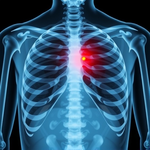
In the evolving landscape of radiation oncology, proton beam therapy (PBT) has emerged as a cutting-edge modality for targeting lung cancers with precision, hoping to minimize damage to surrounding healthy tissues. However, a recent comprehensive retrospective study published in BMC Cancer raises critical awareness about an underappreciated adverse effect: radiation-induced rib fractures (RIRFs) following PBT in patients with stage I non-small cell lung cancer (NSCLC). This investigation delves deeply into the clinical, anatomical, and dosimetric parameters that predispose some patients to this painful complication, revealing complex interplays between radiation dose distribution and individual skeletal factors.
Radiation-induced rib fractures represent a growing concern in thoracic radiotherapy due to the increasing use of hypofractionated regimens, where higher doses per fraction are delivered to reduce treatment times and potentially improve tumor control. While hypofractionation has demonstrated therapeutic benefits, it also amplifies the biological impact on non-target tissues. Particularly with proton and carbon-ion therapies, whose physical dose distributions differ substantially from conventional photon beams, the characterization and mitigation of toxicities such as RIRFs remain inadequately elucidated. This study pioneers an in-depth examination of these risks specifically within the context of passive-scattering proton beam therapy.
The research team conducted a meticulous retrospective analysis of 85 patients afflicted with stage I NSCLC, all treated with a consistent proton dose of 66–70 Gy (relative biological effectiveness, RBE) administered in 10 fractions. A stringent follow-up period of no less than 36 months was enforced to capture late-onset rib fractures, which may often remain asymptomatic and thus undetected without thorough imaging surveillance. This extended observation window is vital, as radiation-induced bone injuries can manifest long after therapy completion, obscuring direct causal links in shorter studies.
Statistical scrutiny encompassed both Kaplan–Meier survival analyses for fracture-free intervals and Cox proportional hazards models to identify robust predictors of RIRFs. Notably, out of 85 patients, 55 (64.7%) experienced at least one rib fracture, culminating in a staggering total of 116 fractured ribs. The cumulative incidence rose steeply over time, with 36.5% and 52.9% of patients sustaining fractures by 2 and 3 years post-treatment, respectively, highlighting this complication’s prevalence and clinical significance.
One of the pivotal findings lies in the dose-response relationship at the level of individual ribs. By focusing on ribs exposed to at least 50 Gy (RBE), the analysis included 224 rib units, encompassing all fractured instances. Higher maximum doses delivered to small volumes within a rib showed a clear association with fracture risk, establishing dosimetry as a critical determinant. The spatial location of the maximum dose also emerged as significant — fractures were more likely when the high dose area corresponded with structurally vulnerable rib regions.
Beyond pure dosimetric factors, the study innovatively incorporated patient-specific skeletal considerations into the risk assessment paradigm. Bone mineral density (BMD), a surrogate marker of overall bone strength, was inversely correlated with fracture incidence. This finding aligns with known biological principles that weaker bones are more susceptible to radiation damage and subsequent mechanical failure. Furthermore, specific anatomical rib segments, such as the first rib, proved particularly vulnerable, potentially attributable to unique biomechanical stresses and vascular supply patterns.
An intriguing clinical variable that modulated fracture risk was the systemic use of corticosteroids. These agents, while therapeutically valuable for many conditions, may exacerbate bone fragility through inhibition of osteoblast activity and promotion of bone resorption. Their identification as an independent risk factor stresses the necessity for multidisciplinary consideration when managing patients undergoing radiotherapy.
The median latency period from treatment to fracture was approximately 23.5 months, with occurrences spanning from as early as five months to as late as over five years post-therapy. This wide temporal range emphasizes the need for long-term, possibly lifelong, surveillance of patients receiving proton therapy to promptly identify and manage late skeletal toxicities.
This study’s findings deliver crucial insights that could guide future radiotherapy planning and patient management. Incorporating detailed dose constraints to safeguard rib integrity, especially limiting high-dose “hot spots” in vulnerable anatomical locations, may reduce RIRF risk. Simultaneously, pre-treatment assessment of bone health, potentially including dual-energy X-ray absorptiometry (DXA) scans to quantify BMD, could stratify patients based on susceptibility, enabling tailored protective strategies.
Moreover, the interplay of corticosteroid use with radiation effects on bone underscores a pressing need for heightened vigilance in patients requiring such medications concurrent with or following radiotherapy. Inter-disciplinary collaboration with endocrinologists or bone metabolism specialists might be prudent to optimize bone-preserving interventions and monitor fracture risk.
These revelations also prompt a broader reflection on radiation toxicity paradigms in proton therapy. Despite protons’ theoretical advantage in sparing healthy tissues due to their characteristic Bragg peak, localized dose escalations remain a double-edged sword, harboring potential for unintended collateral damage. The complex biomechanical milieu of ribs, with varying cortical thickness, marrow composition, and loading patterns, demands an individualized approach in dose sculpting and risk evaluation.
The study’s retrospective design, though informative, invites further prospective investigations with larger cohorts and incorporation of advanced imaging biomarkers to refine predictive models. Future research might also explore protective agents or rehabilitative modalities that could mitigate or ameliorate radiation-induced bone injuries.
Ultimately, this seminal work reverberates beyond NSCLC radiotherapy, encouraging cancer care teams to integrate skeletal health considerations when devising treatment plans involving hypofractionated regimens and charged-particle therapies. Enhancing patient quality of life through minimization of painful, debilitating rib fractures represents a pivotal frontier in the ongoing evolution of oncologic precision medicine.
In summary, the determination of multifaceted risk factors—including dosimetric parameters, anatomical nuances, bone mineral density, and corticosteroid use—opens avenues for more comprehensive and personalized radiation oncology strategies. These insights aim to optimize the therapeutic index of proton beam therapy, maximizing tumor eradication while safeguarding patients from the insidious sequelae of skeletal complications.
As proton therapy continues to gain traction worldwide, integrating these findings into clinical protocols promises not only enhanced safety but also a deeper understanding of radiobiological interactions at the bone-tissue interface. This foundational knowledge lays the groundwork for future innovations that could revolutionize how radiation-induced toxicities are predicted, prevented, and managed in thoracic oncology.
By bridging clinical observations with rigorous dosimetric analysis, this investigation exemplifies the power of multidisciplinary research in advancing the frontiers of radiation medicine. As we stride toward increasingly precise and patient-tailored cancer treatments, acknowledging and addressing the subtle yet impactful risks such as radiation-induced rib fractures will be paramount in delivering truly holistic care.
Subject of Research: Risk factors for radiation-induced rib fractures following proton beam therapy in stage I non-small cell lung cancer.
Article Title: Risk factors for radiation-induced rib fractures following proton beam therapy for stage I non-small cell lung cancer: a retrospective study.
Article References:
Kondo, N., Yoshiura, T., Kakinohana, Y. et al. Risk factors for radiation-induced rib fractures following proton beam therapy for stage I non-small cell lung cancer: a retrospective study. BMC Cancer 25, 682 (2025). https://doi.org/10.1186/s12885-025-14047-6
Image Credits: Scienmag.com
DOI: https://doi.org/10.1186/s12885-025-14047-6
Tags: dosimetric parameters in cancer treatmenthypofractionated radiation therapyminimizing radiation damagenon-small cell lung cancer treatmentpatient safety in cancer treatmentproton beam therapy complicationsproton therapy dose distributionradiation oncology advancementsradiation-induced rib fracturesretrospective study on cancer patientsskeletal factors in radiation therapythoracic radiotherapy risks


