Enzyme- and fermentation-enriched rice bran extracts reduced streptozotocin-induced injury in two pancreatic beta-cell lines and improved several diabetes-related outcomes in a chemically induced mouse model. The study tested extracts with different ferulic acid and γ-oryzanol profiles, finding that processing method and composition affected which cellular and metabolic outcomes improved. These were laboratory and animal findings; the experiments did not test treatment in people, and the mouse model did not demonstrate autoimmune beta-cell destruction or the insulin deficiency characteristic of human type 1 diabetes.
Rice bran is a milling by-product containing carbohydrates, proteins, dietary fiber and bioactive compounds such as γ-oryzanol, phenolic compounds, tocopherols, tocotrienols and phytosterols. Its bulk volume, low packing density, transportation costs and susceptibility to rancidity have limited its use as a high-value material. The researchers prepared seven extracts: conventional aqueous and ethanolic extracts, an enzyme-treated ferulic acid-enhanced preparation, a solid-state-fermented γ-oryzanol-enhanced preparation, mixtures of conventional extracts, mixtures of enhanced extracts and an additional mixed-fermented preparation. High-performance liquid chromatography confirmed that processing altered their chemical composition. The enzyme-treated extract contained 95.28 mg of ferulic acid per 100 g, compared with 29.62 mg in the conventional aqueous extract, while the solid-state-fermented extract contained 4,841 mg of γ-oryzanol per 100 g, compared with 2,057 mg in the conventional ethanolic extract.
For the cellular experiments, the team used NIT-1 cells, derived from a mouse insulinoma, and HIT-T15 cells, derived from hamster pancreatic islet cells. A dose-ranging analysis found that approximately half of NIT-1 cells were nonviable after exposure to 0.4 mM streptozotocin, or STZ, whereas the corresponding concentration for HIT-T15 cells was 0.5 mM. The researchers selected 0.35 mM STZ for subsequent work; at that concentration, viability fell to 72% in NIT-1 cells and 81% in HIT-T15 cells. Co-treatment with 10 micrograms per milliliter of extract generally improved viability, but the results differed by preparation and cell line. GO, FA+GO and FAenh+GOenh significantly improved viability in NIT-1 cells, while the fully fermented mixture did so in HIT-T15 cells. Other extracts had nonsignificant effects, and several further reduced viability in HIT-T15 cells.
Crystal violet staining broadly reflected the MTT viability results, showing stronger staining after STZ injury when extracts were added than with STZ alone, although the experiment did not directly quantify cell attachment. Protection also was not consistently stronger at higher extract concentrations. Tests across 1, 2, 5, 10, 20 and 50 micrograms per milliliter showed that some preparations added little protection at higher doses or became cytotoxic under STZ challenge. The researchers therefore used 10 micrograms per milliliter as a shared concentration for mechanistic experiments while noting that it was not the optimal concentration for every extract. This variability also means the preparations should not be treated as chemically interchangeable interventions: their protection depended on composition, dose and experimental model.
The extracts influenced some of the cellular disturbances associated with STZ, although the clearest effects differed between the cell lines. STZ increased intracellular reactive oxygen species to approximately 120% of the untreated NIT-1 control. Co-treatment with the conventional γ-oryzanol extract, the solid-state-fermented γ-oryzanol extract, the unfermented combined enhanced extract or the mixed fermented extract significantly reduced that signal toward or below control levels. The effect was less clear in HIT-T15 cells, where STZ caused only a small, statistically nonsignificant increase in reactive oxygen species. STZ reduced cytosolic calcium to 64% of control in NIT-1 cells and 66% in HIT-T15 cells. Several extracts significantly restored calcium in HIT-T15 cells, while most produced only nonsignificant trends in NIT-1 cells. The divergence should not be attributed solely to species: the cell lines also differ in genetic background, immortalization history and culture-dependent functional characteristics.
Gene and protein measurements provided partial evidence about possible cellular mechanisms. STZ reduced messenger RNA expression of the mitochondrial fusion regulators mfn1 and opa1 and the anti-apoptotic gene bcl2, and selected extracts partially restored their expression. At the protein level, the solid-state-fermented γ-oryzanol extract significantly restored GLUT1 and PDX1 in NIT-1 cells, while the mixed fermented extract increased GLUT4. In HIT-T15 cells, the conventional ferulic acid extract significantly restored GLUT1, but most changes involving GLUT4 and PDX1 were not statistically significant. The results are consistent with partial preservation of glucose-transport and beta-cell regulatory proteins under chemical stress. They do not establish a complete protective pathway, because the researchers did not directly measure glucose-stimulated insulin secretion, mitochondrial function, apoptosis, beta-cell mass or pancreatic insulin content.
The mouse experiment used five-week-old male C57BL/6N mice. Repeated low-dose STZ initially failed to produce sufficiently stable and consistent hyperglycemia, so the investigators administered an additional 150 mg/kg dose on day 29. On day 35, animals with fasting blood glucose of at least 150 mg/dL were classified as diabetic and allocated non-randomly to a basal-diet control group or diets containing 0.5% rice bran extract. Each group contained eight mice, and dietary treatment continued for 10 weeks. Treated animals consumed less water than untreated diabetic mice, consistent with partial attenuation of excessive drinking, while their fasting glucose declined progressively over time. Oral glucose tolerance also improved, including a significant reduction in overall glucose excursion compared with untreated diabetic mice. Body-weight differences were not statistically significant, and food-intake measurements did not suggest that reduced eating explained the metabolic changes.
Tissue examinations indicated that the supplements also mitigated some pathological changes caused by STZ. Untreated diabetic mice accumulated lipid droplets in the liver and had enlarged pancreatic acinar cells with cytoplasmic vacuolization. Most supplemented groups showed less hepatic lipid accumulation, and liver tissue from the mixed fermented group closely resembled that of naïve controls. Pancreatic islet number and appearance in treated groups moved toward control values, while vacuolization and acinar-cell enlargement diminished. These findings support attenuation of general islet and exocrine pancreatic injury, but the study counted islets without measuring total islet area, beta-cell mass or pancreatic insulin content. Its model was therefore diabetes-like in terms of chemically induced hyperglycemia and tissue injury, but the study did not establish autoimmune beta-cell destruction or insulin deficiency.
Other metabolic measures changed alongside the improved glucose control. Plasma GLP-1 was approximately 2.3-fold higher in diabetic mice than in naïve controls but generally declined toward normal values in the treatment groups. The ferulic acid-enhanced extract was an exception, increasing GLP-1 further. The authors associated this pattern with previously reported inhibition of DPP-4-mediated GLP-1 degradation by ferulic acid, but that mechanism was not directly tested. Epididymal white adipose tissue had more than a 50% lower weight and gross size in diabetic animals; supplemented mice gained approximately 6 to 20 mg relative to untreated diabetic mice. Plasma insulin was unexpectedly higher in the diabetic group than in naïve controls and decreased toward the control level after supplementation. Possible explanations include secretion by residual beta cells or altered insulin clearance, but neither was experimentally examined in this study.
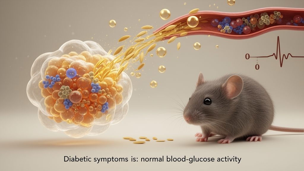
Overall, the experiments indicate that selected processed rice bran materials can attenuate STZ-associated injury in beta-cell models and partially improve hyperglycemia, excessive drinking, glucose tolerance, liver histology, pancreatic architecture, GLP-1 levels and adipose-tissue mass in a mouse model. No single preparation was uniformly best across every outcome: the enzyme-enhanced, γ-oryzanol-enhanced, combined and mixed fermented extracts had distinct effects under particular conditions. Important questions remain, including which constituents were responsible, how the extracts were absorbed and metabolized, and whether their composition could be standardized for longer-term use. The results identify candidate materials and processing strategies for further preclinical investigation, but they do not establish rice bran extracts as a treatment for diabetes in humans.
Subject of Research: Preclinical testing of processed rice bran extracts in STZ-induced beta-cell injury and diabetic mouse models
Article Title: Enhanced rice bran extracts attenuate STZ-induced β-cell injury and diabetes-related metabolic disturbances
Article References: Song, J., Hong, T., Kim, M., Kim, G., Kim, Y., Kim, S. H., Song, G., Park, S., & Lim, W. (2026). Enhanced rice bran extracts attenuate STZ-induced β-cell injury and diabetes-related metabolic disturbances. Journal of Agriculture and Food Research, Article 103377. https://doi.org/10.1016/j.jafr.2026.103377
Image Credits: AI Generated
DOI: 10.1016/j.jafr.2026.103377
Keywords: rice bran, streptozotocin, pancreatic beta cells, ferulic acid, gamma-oryzanol, fermentation, oxidative stress, glucose tolerance, diabetic mice, rice bran extracts, Enhanced, rice
News Source: Alan Morgan. (October 22, 2026). Rice bran extracts ease STZ injury in cells and diabetic symptoms in mice. Scienmag.