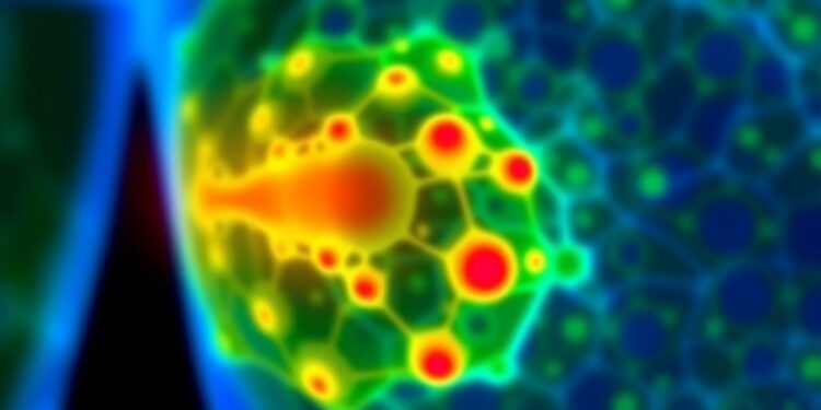
Researchers at the Technion – Israel Institute of Technology have achieved a remarkable breakthrough in the field of biomedical science, particularly in the way we can observe and interact with molecular processes within tissue. Their innovative technology, recently published in the eminent journal Advanced Materials, offers a fresh approach to understanding health and diseases at the molecular level. This pioneering method focuses on monitoring changes within organoids—three-dimensional cell cultures that mirror the structure and functionality of actual organs. By utilizing chemical tomography, this new technique promises to redefine our capacity for early disease detection and personalized medicine, leading the way for future innovations in healthcare.
Organoids serve as a vital tool for scientists, providing a closer representation of human tissues than traditional two-dimensional cell cultures. Their ability to replicate the intricate behaviors of human organs makes them exceptional models for investigating various diseases and testing treatments. However, one of the significant hurdles researchers face with organoids is the challenge of monitoring the internal processes effectively. Existing techniques are often either too costly, destructive to the tissue, or incapable of providing detailed insights into deeper tissue layers. The team at the Technion has successfully devised a method that tackles these limitations, allowing them to observe dynamic changes in organoids without causing damage, all while keeping costs manageable.
The innovation hinges on the analysis of volatile organic compounds (VOCs), which are small molecules emitted from tissues and detected in biological fluids such as breath and sweat. These compounds act as biological markers, offering crucial information about the underlying processes occurring within the tissue. Prof. Hossam Haick, a leading expert on VOCs and their implications for disease detection, highlights the transformative potential of this research. The study revealed significant insights into breast tissue transformation, showcasing how monitoring VOCs can unveil essential genomic and protein alterations associated with cancer.
One standout feature of this breakthrough is its real-time monitoring capability. Unlike traditional methods that provide static snapshots of organoid states, the chemical tomography technique allows researchers to observe how these organoids change over time. They can track cancer progression through various stages, better understand the underlying biology of the disease, and even map intricate biochemical pathways and metabolic markers responsible for cancer development. The identification of six distinct biochemical pathways that yield twelve different types of VOCs stands as a testament to the method’s power in elucidating complex biological systems.
The implications of this research extend far beyond cancer detection. According to Prof. Haick, their methodology has potential applications in diagnosing various health conditions affecting organs like the kidneys, brain, and liver. The system’s design includes the possibility of real-time transmission of health data to external monitoring systems through antennas, enabling continuous tracking of tissue health and providing early warning signs of potential diseases. This capability marks a significant advancement in incorporating artificial intelligence into healthcare, steering us closer to truly personalized medicine tailored to individual patient needs.
Moreover, this research resonates with the broader goal of integrating technological advancements with traditional healthcare practices. The ability to non-invasively monitor molecular processes could facilitate more timely and accurate diagnoses, drastically improving patient outcomes. The collaboration between multiple research institutes, including the University of Haifa, enhances the interdisciplinary nature of this project, empowering researchers to pool their expertise and tackle complex health challenges more effectively.
As this new method garners attention in the scientific community, it raises exciting possibilities for future innovations. The marriage of biotechnology, AI, and molecular imaging signifies a pivotal shift in how we approach diagnostics and treatment in modern medicine. As healthcare professionals adopt these advanced tools, they stand to revolutionize patient care, leading to more precise interventions and less reliance on costly and invasive procedures.
The recognition of the study by the prestigious journal Advanced Materials and the backing from organizations like The Zimin Foundation and The European Research Council underscores the significance of this work. Such validation from reputable sources adds credibility to the researchers’ findings and indicates a shared belief in the potential impact of their contributions to personalized healthcare and disease detection.
The advent of this new technique not only promises to elevate the quality of cancer research but holds profound implications for comprehensive healthcare improvements. As researchers continue to explore and refine this technology, we may find ourselves embarking on a new era in medicine, where real-time monitoring and early intervention become standard practice, fundamentally changing our approach to treating diseases and managing health.
This pioneering study accentuates the clear necessity for continued investment in research and development. Encouraging collaboration among scientists across various fields will be essential in harnessing the full capabilities of modern technologies, ensuring that the potential of innovations is realized in tangible ways that positively impact patients’ lives. The Technion team’s findings set a new standard for organoid research, offering an optimistic trajectory for the future of medical diagnostics and treatment, highlighting the interconnectedness of technology and health.
In summary, this breakthrough research from the Technion offers an exciting glimpse into the future of health diagnostics. The successful integration of chemical tomography with AI to detect VOCs has pushed the boundaries of what is possible in medical science. As we look to the future, there is little doubt that this transformative approach has the potential to change how we understand, diagnose, and treat diseases, opening up a wealth of new possibilities that will benefit countless individuals around the world.
Subject of Research: Lab-produced tissue samples
Article Title: Chemical Tomography of Cancer Organoids and Cyto-Proteo-Genomic Development Stages Through Chemical Communication Signals
News Publication Date: 11-Feb-2025
Web References: 10.1002/adma.202413017
References: N/A
Image Credits: N/A
Keywords: Cancer research, Chemical tomography, Personalized medicine, VOC analysis, Graphene sensors, AI in healthcare, Biomedical innovation
Tags: advanced materials in biomedical researchbiomedical breakthroughs at Technionchemical tomography for disease detectiondeep tissue molecular processesearly disease detection techniquesinnovative healthcare solutionsmolecular level health insightsmonitoring organoid internal processesnon-invasive tissue monitoringorganoid technology in healthcarepersonalized medicine advancementsthree-dimensional cell cultures