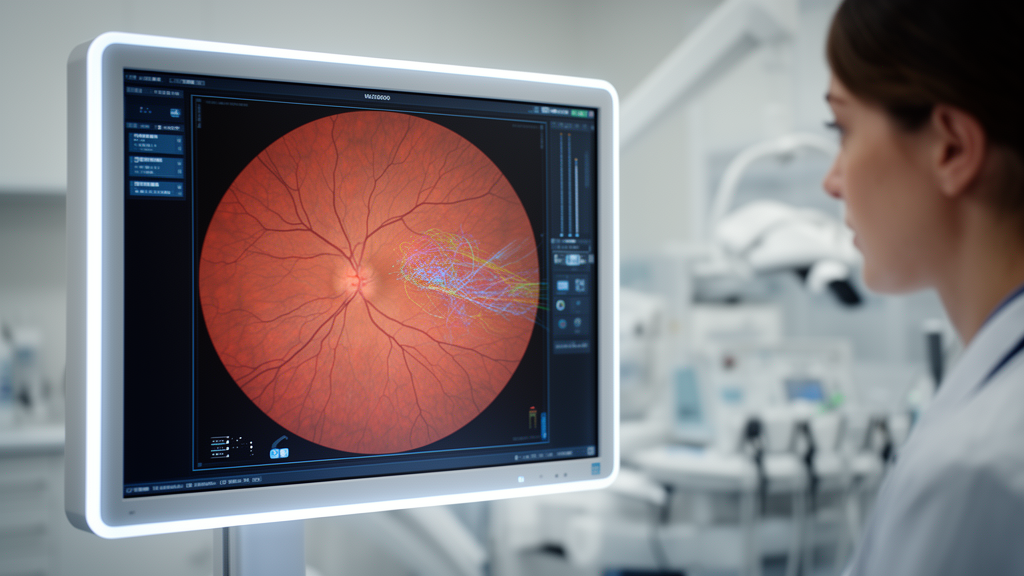
Retinal ischemia serves as a common pathological thread linking several vision-threatening ocular disorders, yet high diagnostic accuracy among artificial intelligence models evaluated for these conditions does not automatically guarantee clinical utility or seamless integration into real-world healthcare pathways. In a narrative review published in Frontiers in Artificial Intelligence, researchers led by Fapianey J. Alexandre and colleagues at Meharry Medical College critically evaluated eighteen primary studies examining machine learning and deep learning applications across retinopathy of prematurity, diabetic retinopathy, and retinal vein occlusion. Retinal tissue ranks among the most metabolically demanding structures in the human body, relying on uninterrupted oxygen delivery to sustain normal visual function. Impairment of microvascular perfusion triggers tissue hypoxia, capillary nonperfusion, vascular leakage, inflammatory cascades, and compensatory upregulation of vascular endothelial growth factor, culminating in pathological neovascularization. Although incomplete vascular development drives ischemic injury in retinopathy of prematurity, progressive microvascular damage characterizes diabetic retinopathy, and retinal venous outflow obstruction defines retinal vein occlusion, automated analysis of retinal imaging modalities has emerged as a promising tool for identifying and characterizing these shared manifestations of ischemic injury.
To investigate how artificial intelligence models perform across these distinct clinical scenarios, the authors executed structured literature searches in PubMed and Google Scholar through August 2026, retrieving 626 records. After excluding duplicate citations, screening titles and abstracts, and completing full-text eligibility reviews, sixty-four publications were excluded due to review formats, preclinical animal designs, insufficient methodological reporting, or lack of direct clinical relevance. The resulting core evidence base comprised eighteen unique primary studies that represented twenty-six distinct outcome-level records spanning diagnostic disease characterization, longitudinal disease quantification, predictive modeling, and operational clinical workflow deployment. The reviewed algorithms evaluated diverse imaging inputs, including wide-angle fundus photographs, conventional color fundus photographs, optical coherence tomography, optical coherence tomography angiography, and fundus fluorescein angiography. However, the authors emphasized that study identification involved author judgment, exact database queries were not archived, and methodological heterogeneity across the literature prevented quantitative meta-analytic data pooling, requiring findings to be synthesized within their specific clinical contexts.
Diagnostic and disease-characterization applications represented the most extensively investigated area within the literature, demonstrating strong discriminatory power on curated image sets across all three retinal conditions. In retinopathy of prematurity, deep learning models focused heavily on recognizing plus disease, an assessment traditionally complicated by subjective clinical interpretation of arterial tortuosity and venous dilation. For instance, the i-ROP deep learning system achieved an area under the receiver operating characteristic curve of 0.98 in development evaluations and demonstrated 93 percent sensitivity and 94 percent specificity on an independent 100-image test set. Another model, ROP.AI, achieved an area under the curve of 0.977 on an external set of ninety images, though specificity declined to 80.7 percent outside the primary development cohort. Diagnostic studies in diabetic retinopathy established even larger algorithmic benchmarks, with deep convolutional neural networks trained on over 128,000 images achieving areas under the curve of 0.991 on EyePACS-1 and 0.990 on Messidor-2 for referable disease, while separate multiethnic evaluations achieved 90.5 percent sensitivity and 91.6 percent specificity across 71,896 validation images.
In retinal vein occlusion, diagnostic research diverged from standardized binary screening classifications toward multi-label subtype categorization, angiographic interpretation, and objective vascular quantification. A neural understanding network distinguished normal retinal photographs from central and branch retinal vein occlusions with 91.1 percent accuracy and 98.3 percent sensitivity, though specificity was lower at 80.3 percent. A Swin Transformer architecture differentiated normal retinas, central vein occlusion, branch vein occlusion, and macular retinal vein occlusion with class-specific areas under the curve spanning 0.900 to 1.000 across a small cohort of 914 fundus photographs. Moving beyond categorical disease labeling, deep learning denoising applied to optical coherence tomography angiography in branch retinal vein occlusion improved the area under the curve for distinguishing moderate from severe macular ischemia from 0.802 to 0.927 for vessel density and from 0.797 to 0.946 for nonperfusion area. Additionally, a multimodal system combining optical coherence tomography, infrared reflectance imaging, and clinical diabetes status differentiated macular edema causes with 95.20 percent accuracy across four diagnostic categories, showing the versatility of automated feature extraction.
Beyond assessing established disease, predictive and longitudinal applications aimed to anticipate future disease onset or track changes over time, though evidence supporting these clinical functions remained considerably more limited. Predictive modeling was identified only in retinopathy of prematurity and retinal vein occlusion within the eighteen primary studies, with no predictive diabetic retinopathy studies meeting inclusion criteria. In retinopathy of prematurity, an investigation combining baseline screening photographs with forty-six clinical demographic variables achieved external areas under the curve of 0.94 for predicting disease occurrence and 0.88 for predicting severity. However, when operating thresholds were calibrated to maximize clinical sensitivity, external specificity dropped precipitously to 7.5 percent for occurrence prediction and 35.3 percent for severity prediction, an operating profile that would generate an overwhelming volume of false-positive alarms in clinical practice. Longitudinal assessment in retinopathy of prematurity was separately illustrated by a deep learning vascular severity score ranging from one to nine, which tracked continuous vascular changes through treatment and regression across 5,255 examinations from 871 infants without prospectively forecasting individual clinical endpoints.
Predicting therapeutic outcomes in retinal vein occlusion similarly revealed preliminary feasibility alongside significant translational hurdles. Investigators evaluating pretreatment fundus fluorescein angiograms with a transfer-learning convolutional neural network named FFA-Net attempted to forecast short-term response to anti-vascular endothelial growth factor injections among 180 patients presenting with branch retinal vein occlusion and macular edema. The model attained an overall accuracy of 88.63 percent, an F1 score of 0.89, a sensitivity of 79.40 percent, a specificity of 71.34 percent, and an area under the curve of 0.71. Although gradient-weighted class activation mapping helped visualize image regions contributing to network outputs, the underlying investigation remained constrained by a single-center retrospective design, brief follow-up periods, modest statistical discrimination, and a lack of independent external cohort validation, underscoring that predicting therapeutic response remains technically demanding.
Translational readiness and operational workflow integration varied widely across the three vascular diseases, with diabetic retinopathy advancing furthest toward real-world deployment. Prospective clinical trials in primary care established the viability of autonomous screening algorithms that operate without immediate specialist overread. A pivotal prospective multicenter trial evaluating the IDx-DR autonomous system across 900 primary-care participants achieved 87.2 percent sensitivity and 90.7 percent specificity for detecting more-than-mild diabetic retinopathy, with an imageability rate of 96.1 percent. Similarly, the EyeArt autonomous system demonstrated 95.5 percent sensitivity and 85.0 percent specificity for more-than-mild disease across 893 analyzable participants in fifteen clinical sites, alongside 95.1 percent sensitivity for vision-threatening retinopathy. Prospective validation in resource-limited environments was corroborated in Zambia, where an algorithm developed abroad yielded 92.25 percent sensitivity and 89.04 percent specificity on 4,504 fundus images, demonstrating that automated image assessment can successfully operate across geographically diverse patient demographics.
Conversely, clinical workflow evaluations in retinopathy of prematurity and retinal vein occlusion have not attained comparable levels of prospective operational deployment. In retinopathy of prematurity, workflow research was restricted to a retrospective evaluation of an automated vascular severity score applied to 613 examinations collected within an operational telemedicine screening network, achieving an area under the curve of 0.99 for identifying referral-requiring disease. While illustrating potential for triage prioritization, the study did not prospectively assess algorithmic referral routing or quantify patient outcomes. In retinal vein occlusion, none of the included investigations examined models prospectively within an intended clinical care setting. The authors noted that eight of the eighteen review studies were limited entirely to internal retrospective evaluations, six attained independent external validation, three completed prospective clinical validation, and only one assessed operational workflow data, illustrating that current literature remains heavily skewed toward early-stage technical development rather than practical deployment.
The review also highlighted technical and human-factor constraints that undermine model generalizability when systems transition from curated benchmark datasets to routine clinical practice. Variations in camera hardware, exposure quality, patient movement, media opacities, and ungradable photographs directly impair diagnostic reliability. Automated quality triage was demonstrated by an image-quality classifier that achieved an area under the curve of 0.965, 93.9 percent sensitivity, and 83.6 percent specificity across 2,109 held-out pediatric images, confirming that quality gating represents an essential operational step before diagnostic inference. Furthermore, common interpretability tools such as saliency maps and Grad-CAM visualizations provide only post-hoc spatial correlations rather than transparent causal explanations of algorithmic reasoning, and distinct explanatory techniques frequently produce conflicting visual outputs. Consequently, human clinician oversight remains mandatory for reconciling discordant outputs, evaluating entire clinical histories, and guiding definitive therapeutic interventions.
Ultimately, the synthesized evidence demonstrates that regulatory authorization and headline area under the curve statistics do not inherently translate into improved downstream clinical care or preserved visual acuity. In the United States, autonomous screening platforms like IDx-DR have obtained marketing clearance through De Novo pathways, while systems such as EyeArt and AEYE-DS received 510(k) clearances, yet real-world deployment data show that technical screening success does not guarantee completed specialist follow-up. For example, a 2026 endocrinology clinic study of AEYE-DS achieved definitive autonomous screening results in 95.7 percent of patients without pharmacological dilation, yet only 44.7 percent of screening-positive patients completed confirmatory ophthalmological examinations at the same institution. Continuous post-market surveillance, rigorous external validation, standardized reporting of statistical uncertainty, and dedicated prospective investigations evaluating referral completion, diagnostic timeliness, treatment burden, and cost-effectiveness will be required before artificial intelligence can safely and effectively optimize patient care across ischemic retinal diseases.
Subject of Research: Translational readiness of artificial intelligence across ischemic retinal diseases
Article Title: Artificial intelligence in ischemic retinopathy: from diagnostic and predictive performance to clinical translation
Article References: Alexandre, F. J., Brokemond, A. J., Amamoo, R., Ajisegiri, S. B., Bailey, B., Martin, P. M., Jadeja, R. N., & Thounaojam, M. C. (2026). Artificial intelligence in ischemic retinopathy: from diagnostic and predictive performance to clinical translation. Frontiers in Artificial Intelligence, 9, Article 1994986. https://doi.org/10.3389/frai.2026.1994986
Image Credits: AI Generated
DOI: 10.3389/frai.2026.1994986
Keywords: artificial intelligence, diabetic retinopathy, retinopathy of prematurity, retinal vein occlusion, ischemic retinopathy, deep learning, fundus photography, optical coherence tomography angiography, fundus fluorescein angiography, autonomous screening, clinical translation, diagnostic performance, treatment response prediction, vascular severity score, image quality assessment, ophthalmology, telemedicine, post-market surveillance
News Source: Blake Davidson. (November 18, 2026). Retinal Artificial Intelligence Demonstrates High Diagnostic Accuracy but Faces Clinical Implementation Barriers. Scienmag.