A new, reproducible mouse calvarial defect model has been successfully established to study cranial osteomyelitis induced by Staphylococcus aureus, providing a versatile platform for exploring the mechanisms of bone infection in the skull. Osteomyelitis, a severe inflammatory bone disease, often necessitates complex surgical debridement and prolonged high-dose antibiotic therapy due to chronic inflammation, tissue necrosis, and vascular compromise that frequently lead to treatment failure. While various animal models have been developed to mimic the diverse etiologies and skeletal manifestations of this infection, researchers have faced consistent challenges in creating a reliable model for cranial osteomyelitis in mice. By utilizing bilateral calvarial defects with controlled bacterial inoculation, this study identifies a specific bacterial inoculum threshold that reliably produces destructive bone infection, potentially enabling future research on craniofacial infection pathways and therapeutic interventions.
Osteomyelitis is generally classified based on its etiology into cases arising from contiguous infection, vascular insufficiency, or hematogenous spread. Staphylococcus aureus remains the most frequent pathogen isolated in clinical bone infections, making it a primary target for experimental modeling in diverse species, including rabbits, rats, and larger mammals. However, rodents like mice offer unique advantages for researchers, such as cost-effectiveness, ease of handling, and the ability to employ transgenic or knockout models to investigate specific molecular signaling pathways or immune responses. Despite these benefits, mice have not been widely adopted for osteomyelitis research, primarily due to technical limitations regarding their small scale, the difficulty of surgical procedures, and the limited success in performing two-stage revision surgeries. Furthermore, existing mouse models have focused almost exclusively on long bones, which undergo endochondral ossification, failing to capture the distinct regenerative environment of the skull, which is formed through intramembranous ossification.
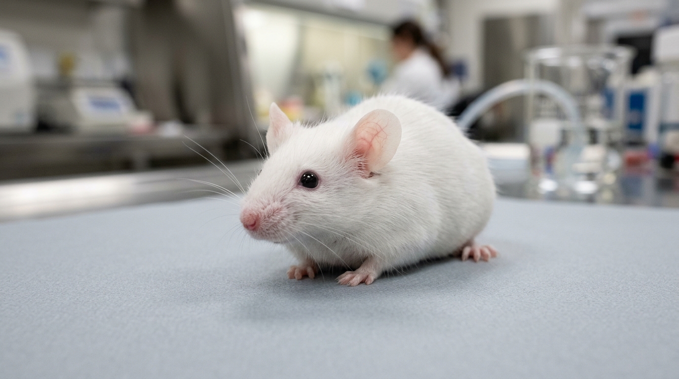
The study design involved 27 male C57BL/6J mice, which were randomly assigned to five groups, including a control group and four experimental cohorts. After an acclimatization period, the mice underwent surgery where bilateral 4-mm diameter calvarial defects were created using a trephine bur. The experimental groups received varying concentrations of S. aureus (ATCC 6538) at doses of 10^5, 10^6, 10^7, and 10^8 colony-forming units (CFU)/mL, while the control group received an inoculation of sterile phosphate-buffered saline (PBS). The animals were housed in an animal biosafety level 2 facility under specific pathogen-free conditions with standardized environmental controls. All experimental procedures were approved by the local institutional animal care and use committee, and all mice were monitored until the scheduled endpoint four weeks post-surgery to observe clinical outcomes and evaluate the success of the infection model.
Micro-computed tomography, known as micro-CT, served as a primary diagnostic tool to assess bone regeneration and structural integrity across the experimental groups. Analysis of the bone volume/total volume (BV/TV) ratio revealed a clear dose-response relationship where higher bacterial concentrations resulted in significantly reduced bone regeneration. Although bone mineral density (BMD) values did not show significant pairwise differences between specific groups, there was a significant linear decreasing trend across the ordered bacterial concentrations, suggesting a progressive reduction in mineralization as the bacterial challenge increased. Additionally, the bony bridging and defect union scores were markedly lower in the 10^8 CFU/mL cohort, reflecting a consistent failure of bone repair in the presence of higher bacterial burdens.
Bacteriological analysis further confirmed the presence and magnitude of the infection in the harvested calvarial bone. Bone samples were processed using an ultrasonic homogenizer and plated to determine the viable bacterial count. The recovery of CFU counts demonstrated that the 10^8 CFU/mL inoculum group achieved significantly higher bacterial burdens compared to the control and the lowest-dose group. While groups inoculated with lower bacterial concentrations did show intermittent bacterial growth, the results were not statistically distinct from the control group, indicating that lower inocula were insufficient to reliably cause the severe infection observed at the 10^8 CFU/mL threshold. This finding serves as a practical, objective benchmark for future studies needing to replicate destructive cranial osteomyelitis in a mouse model.
Histological evaluation provided deeper insights into the pathological features of the induced infection, utilizing a semi-quantitative scoring system to assess tissue-level inflammation, bone necrosis, and the presence of bacterial colonies. Hematoxylin and eosin (H&E) staining of the defect margins revealed connective tissue coverage in all groups, but the outcomes of the infection groups varied significantly. These histological indicators—including the composite score of inflammation, necrosis, and bacterial presence—were consistently and significantly higher in the 10^8 CFU/mL group compared to the control group. These observations corroborate the micro-CT findings, confirming that the model successfully replicates the key histological features associated with cranial osteomyelitis.
The mechanisms underlying the observed bone loss in this model are believed to align with known patterns of osteomyelitis, which often trigger an imbalance in the receptor activator of nuclear factor kappa-B ligand (RANKL) and osteoprotegerin (OPG) signaling pathway. An increased RANKL/OPG ratio is frequently associated with enhanced osteoclast differentiation and activity, which contributes to bone resorption and decreased mineral density during infection. The progressive linear trend observed in the BMD data supports this hypothesis, indicating that the bacteria-driven inflammatory environment directly influences bone turnover and structure. These results suggest that the mouse model is not only useful for confirming the presence of infection but also for probing the molecular mechanisms that drive bone loss within the specialized environment of the calvaria.
The development of this mouse model addresses a long-standing gap in craniofacial research, where the fundamental differences in vascularization and regenerative microenvironments between long bones and the skull have hindered the direct application of existing knowledge. The skull’s unique anatomy and cosmetic considerations necessitate models that accurately reflect these conditions, and the established 10^8 CFU/mL inoculation protocol provides a reliable tool for researchers. Furthermore, the model’s compatibility with transgenic and knockout approaches opens new avenues for studying the immune response and the influence of specific genetic factors in the pathogenesis of skull infections. This platform is anticipated to facilitate preclinical testing of various local and systemic therapeutic strategies targeting bone infections in the craniofacial region.
Despite the successful induction of the model, the study acknowledged certain limitations that remain to be addressed in subsequent research. Additionally, the use of only one strain of S. aureus (ATCC 6538) restricts the immediate generalizability of the inoculum cut-off, as virulence and biofilm-forming capacities differ widely among clinical isolates.
This study represents a significant advancement by demonstrating that a reproducible mouse model of calvarial defect osteomyelitis is feasible and robust when the appropriate bacterial concentration is used. The data provide a foundational protocol that can be leveraged to explore novel therapeutic interventions, including regenerative scaffolds or advanced anti-infective strategies tailored for craniofacial bone infections. By bridging the technical gap in animal modeling, this work sets the stage for more complex experimental designs that incorporate comorbid factors and genetic manipulations. Ultimately, the successful validation of this model strengthens the research toolkit available for addressing the persistent challenge of cranial osteomyelitis, ensuring that future studies can proceed with a standardized, reliable, and cost-effective approach that exploits the unique advantages of the mouse research platform.
Subject of Research: Development of a mouse calvarial defect model for Staphylococcus aureus-induced osteomyelitis.
Article Title: A novel mouse calvarial defect model of osteomyelitis induced by Staphylococcus aureus (ATCC 6538)
Article References: Ko, S., Woo, H. M., Kim, K., & Kim, J. (2026). A novel mouse calvarial defect model of osteomyelitis induced by Staphylococcus aureus (ATCC 6538). Laboratory Animal Research, 42(1), Article 33. https://doi.org/10.1186/s42826-026-00289-3
Image Credits: AI Generated
DOI: 10.1186/s42826-026-00289-3
Keywords: Osteomyelitis, Mouse Model, Calvarial Defect, Staphylococcus aureus, Bone Infection, Craniofacial Surgery, Bone Regeneration, Preclinical Research, Cranial Osteomyelitis, Bone Mineral Density, Histopathology, Animal Biosafety, Intramembranous Ossification, Infectious Diseases, Bone Necrosis
News Source: Kristina Jarvis. (November 17, 2026). Researchers Establish Novel Mouse Model for Cranial Bone Infection Study. Scienmag.