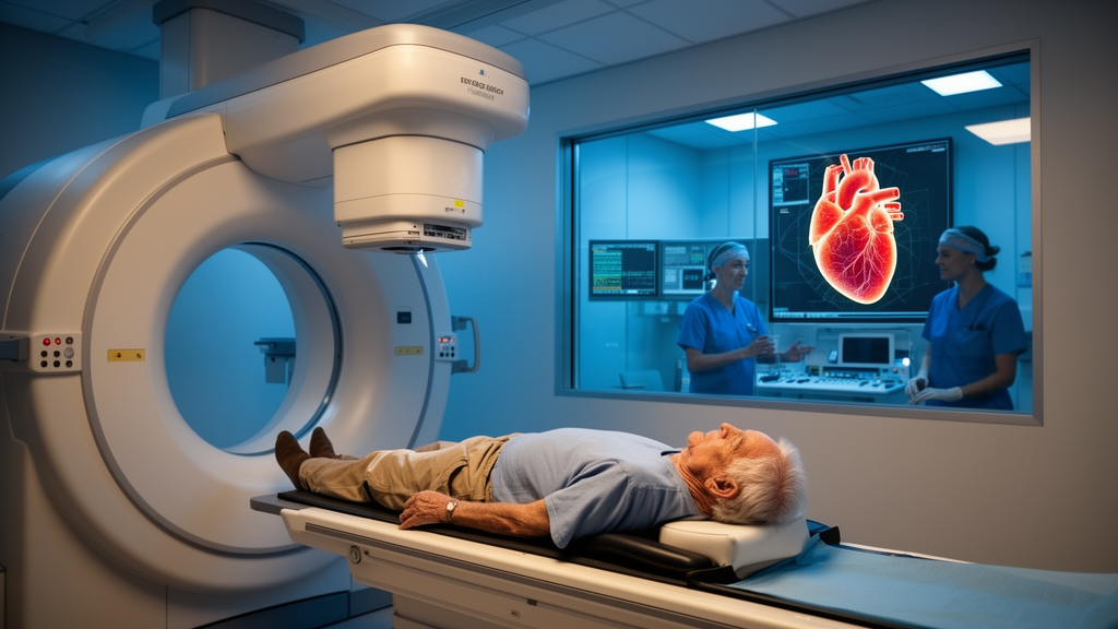
For patients whose hearts are trapped in a relentless rhythm of dangerous electrical storms, a treatment borrowed from cancer therapy is quietly rewriting the rules of cardiac electrophysiology. Stereotactic arrhythmia radioablation, known as STAR, delivers a precisely focused, high dose of radiation to the arrhythmogenic regions of the heart without a single incision or catheter. A new review published in Frontiers in Oncology by researchers at Weill Cornell Medicine synthesizes the radiobiology, clinical evidence, and emerging applications of this technique, and its central message is striking: the treatment appears to work through mechanisms that scientists did not originally predict, and those mechanisms are now reshaping how doses are chosen and how future trials are designed.
STAR was conceived as a non-invasive alternative to catheter ablation, the standard procedure for drug-refractory ventricular tachycardia, in which radiofrequency energy or freezing is used to destroy the cardiac tissue that sustains abnormal rhythms. Catheter ablation has real limitations: it often cannot create deep or transmural lesions across thickened arrhythmogenic substrates, and it cannot safely reach certain epicardial targets, particularly in patients with dense adhesions from previous cardiac surgery or prior mediastinal radiotherapy. STAR sidesteps these obstacles by targeting the myocardium from outside the body with image-guided, high-precision radiation, drawing on decades of experience with stereotactic body radiotherapy and radiosurgery in oncology.
The approach requires a genuinely multidisciplinary team of radiation oncologists, cardiologists, electrophysiologists, and medical physicists, and its implementation still varies considerably between centers. The original biological rationale was straightforward. Animal studies had shown that very high radiation doses, between 40 and 160 Gy, could induce transmural myocardial fibrosis and even atrioventricular conduction block, while substantially lower doses failed to produce comparable structural changes.
On that basis, the first clinical STAR treatments used a single 25 Gy fraction, with the expectation that radiation would reproduce the scar-forming effects of thermal ablation and permanently interrupt re-entrant circuits. Clinical reality, however, told a different story. Patients frequently experienced a marked reduction in ventricular arrhythmias within days to weeks of treatment, a time course far too rapid to be explained by the slow maturation of fibrotic scar tissue.
That discrepancy prompted a search for faster-acting mechanisms, and experimental work has now identified what may be the dominant early effect: electrophysiological reprogramming. Using murine models and explanted human cardiac tissue from patients previously treated with STAR, researchers demonstrated persistent upregulation of two key cardiac conduction proteins, connexin-43, the principal gap-junction protein in ventricular cardiomyocytes, and NaV1.5, the predominant voltage-gated sodium channel responsible for rapid depolarization. These molecular changes were accompanied by accelerated ventricular conduction.
By increasing conduction velocity and reducing electrical heterogeneity within the irradiated substrate, radiation may disrupt the conditions required for sustained re-entry, suppressing arrhythmias before any structural scar forms. Critically, this response appears dose-dependent: single fractions of 15 and 20 Gy produced similar electrophysiological changes, whereas doses of 10 Gy or lower failed to do so. The delayed phase of the radiation response tells a more cautionary story.
Weeks to months after irradiation, the myocardium undergoes vascular injury, inflammation, extracellular-matrix remodeling, fibroblast activation, and variable degrees of fibrosis. Late fibrosis may become apparent roughly three to four months after cardiac irradiation, and an in vivo electroanatomical mapping study performed eight months after STAR reported reduced bipolar voltage in regions receiving at least 15 Gy, suggesting progressive structural remodeling. These delayed changes may contribute to durable substrate modification, but they may also underlie long-term toxicity.
The review highlights particular concern about the vasculature: radiation is an established risk factor for cardiovascular morbidity following thoracic radiotherapy, single-fraction doses above 10 Gy are generally considered capable of inducing substantial vascular injury, and a recent preclinical study showed that a single 16 Gy fraction to different cardiac regions in mice produced structural and functional abnormalities, with the most persistent alterations after irradiation of the cardiac base. This matters enormously for STAR patients, who frequently have ischemic cardiomyopathy or diffuse coronary artery disease with already compromised vascular reserve. These biological uncertainties have direct clinical consequences.
If electrophysiological reprogramming drives the therapeutic benefit while vascular injury and fibrosis drive late toxicity, the optimal dose may not coincide with the traditionally assumed ablative dose. Fundamental radiobiological parameters remain unknown, including the alpha/beta ratio of cardiac tissue after single-fraction irradiation, which limits accurate estimation of radiation tolerance using the linear-quadratic model; while an alpha/beta value of approximately 3 Gy is commonly assumed for late-responding tissues, experimental studies in rat hearts suggest values potentially approaching 1 Gy. Fractionation, which allows repair of sublethal damage between fractions while maintaining biological effectiveness, could theoretically reduce late vascular toxicity, but whether these principles translate to cardiac radioablation is an open question that the field has only begun to address systematically.
The clinical evidence, while encouraging, remains limited in scale. The largest dataset comes from the prospective, multicenter STOPSTORM.eu registry, which included 193 patients with refractory ventricular tachycardia treated across 28 European centers, with a median dose of 25 Gy in a single fraction. Among the 107 patients with at least six months of follow-up, the median reduction in ventricular tachycardia burden was 80 percent compared with the six months before treatment, and 72 percent of evaluable patients remained free of implantable cardioverter-defibrillator shocks during the first six months.
Yet ventricular tachycardia recurred in 55 percent of the overall cohort, and survival probabilities of 88 percent at six months and 77 percent at twelve months reflect the advanced underlying cardiac disease in this population. Twelve serious adverse events were judged possibly or probably related to treatment, including pericardial effusion, coronary events, and early post-treatment arrhythmia, but no deaths were considered treatment-related. Earlier single-arm trials tell a similar story with longer horizons.
The ENCORE-VT phase I/II trial treated 19 patients using non-invasive electrophysiological mapping and reported a 94 percent reduction in aggregate ventricular tachycardia episodes, from 1,778 episodes in the six months before treatment to 111 in the six months after, with the median episode count falling from 199 to 3. At a median follow-up of 24.1 months, however, overall survival had fallen to 58 percent, with eight deaths, four considered possibly treatment-related, and late serious adverse events included a grade 3 pericardial effusion and a grade 4 gastro-pericardial fistula requiring surgical repair. A systematic review and meta-analysis by the STOPSTORM consortium identified ten prospective trials encompassing 82 patients treated between 2016 and 2022, showing reductions of at least 50 percent in ventricular tachycardia burden in 90 percent of evaluable patients and moderate toxicity of 10 percent grade 3 or higher adverse events at 90 days, but one-year recurrence-free survival of only 21 percent.
Even the definition of success is contested: while most studies applied a six-week blanking period, current EHRA/HRS consensus recommends reporting all ventricular arrhythmic events without one, and in the STOPSTORM registry 52 percent of all episodes in the first six months occurred within the first six weeks. Target definition remains one of the field's most stubborn challenges, because the arrhythmogenic substrate is defined by electrical properties rather than visible anatomy. Clinicians therefore rely on surrogates: late gadolinium enhancement cardiac magnetic resonance to identify scar core and border-zone regions, invasive three-dimensional electroanatomic mapping, and non-invasive electrocardiographic imaging, which offers lower spatial resolution.
Dedicated segmentation platforms such as inHEART now integrate multimodal imaging with electrophysiological data to generate three-dimensional representations of the substrate for treatment planning. At Weill Cornell Medicine, five patients treated between September 2019 and April 2023 with volumetric modulated arc therapy delivering 25 Gy in one fraction experienced no acute treatment-related adverse events, with arrhythmia significantly reduced or abrogated at a median of 24 weeks; one patient remained alive and free of ventricular tachycardia events at 50 months. Building on that experience, the institution has opened a randomized phase I/II trial investigating progressive single-fraction dose reduction to 20, 15, and 10 Gy alongside biologically equivalent three-fraction regimens of 36, 30, and 24 Gy, the first prospective effort to optimize the therapeutic ratio of the technique.
Technology is advancing on parallel fronts. Dosimetric comparisons show that CyberKnife plans achieve steeper dose gradients than conventional linear accelerator plans, with a lower gradient measure of 1.00 versus 1.17 centimeters, but require substantially longer treatment times of 119.2 versus 22.4 minutes. MRI-guided linear accelerators promise real-time soft-tissue visualization and online adaptation, while proton therapy, still investigational, reduced mean heart dose from 5.5 to 3.6 Gy and mean lung dose from 1.2 to 0.6 Gy in a dosimetric analysis of 34 patients.
Beyond ventricular tachycardia, early experience in paroxysmal atrial fibrillation has shown that a single 25 Gy fraction to the pulmonary vein antra can achieve durable pulmonary vein isolation confirmed by invasive mapping up to twelve months after treatment, though the thin atrial wall, the nearby right phrenic nerve, esophagus, and coronary arteries make planning treacherous. Randomized trials including RADIATE-VT and CARA-VT are now comparing STAR directly against repeat catheter ablation, and their results will determine whether radiation, once a cancer therapy, earns a permanent place in the cardiology suite.
Subject of Research: Radiobiology, clinical evidence, and emerging applications of stereotactic arrhythmia radioablation for refractory cardiac arrhythmias
Article Title: Stereotactic arrhythmia radioablation: radiobiology, clinical evidence, and emerging applications
Article References: Gregucci, F., Ng, J., Cheung, J. W., Thomas, G., Liu, C. F., Markowitz, S. M., Talebi, F., Roth, B., Patel, M., Teal, A., Muller, L., Zhou, X. K., Pennell, R., & Formenti, S. C. (2026). Stereotactic arrhythmia radioablation: radiobiology, clinical evidence, and emerging applications. Frontiers in Oncology, 16, Article 1953364. https://doi.org/10.3389/fonc.2026.1953364
Image Credits: AI Generated
DOI: 10.3389/fonc.2026.1953364
Keywords: stereotactic arrhythmia radioablation, ventricular tachycardia, cardiac radioablation, radiobiology, electrophysiological reprogramming, connexin-43, catheter ablation, atrial fibrillation, pulmonary vein isolation, radiation toxicity, dose fractionation, STOPSTORM registry
News Source: Ophelia Keating. (October 26, 2026). Research report: Stereotactic arrhythmia radioablation: radiobiology, clinical evidence, and emerging applications. Scienmag.