Physicians treating a 63-year-old woman with recurrent coughing of blood have reported a technique that may widen the options for removing calcified airway stones in patients with low platelet counts. The woman, who carried a history of pulmonary tuberculosis, was found to have a broncholith, a calcified lesion lodged in a distal branch of the right upper lobe bronchus. Her preoperative platelet count was 49 × 10^9/L, below the 50 × 10^9/L mark.
Rather than pulling the stone out whole, the team used a 1.1 mm disposable cryoprobe that freezes onto the stone, combined with biopsy forceps, to break it apart and extract it in pieces. The case, published as a clinical report, suggests that a platelet count below 50 × 10^9/L is not a contraindication for broncholith removal, and that cryoprobe-assisted fragmentation under flexible bronchoscopy may be a feasible therapeutic option for selected patients with transbronchial broncholiths. Broncholithiasis, the condition in which such calcified stones form within the airways, is a relatively rare respiratory disorder.
According to the report, broncholiths are often caused by tuberculosis and histoplasmosis infections. Some patients may be asymptomatic, but when symptoms such as haemoptysis, recurrent infections or airway obstruction occur, broncholith removal may be considered. The patient in this report had been admitted to hospital twice for haemoptysis.
During her first hospitalization, laboratory investigations revealed a platelet count of 66 × 10^9/L, and a computed tomography scan showed a calcified lesion in the distal anterior segmental bronchus of the right upper lobe. She was advised to undergo further testing to determine the cause of her thrombocytopenia but refused, and she was discharged after receiving conservative management. Six months later she was readmitted with coughing of blood again.
This time, laboratory investigations showed a platelet count of 56 × 10^9/L, and a contrast-enhanced CT scan revealed a 9 × 8 mm calcified lesion in the same distal segmental bronchus, sitting adjacent to several pulmonary vascular branches. A preoperative platelet count taken shortly before the procedure was 49 × 10^9/L. Given the combination of a firmly anchored stone, nearby pulmonary vascular branches and the low platelet count, the hospital convened a multidisciplinary assessment before proceeding.
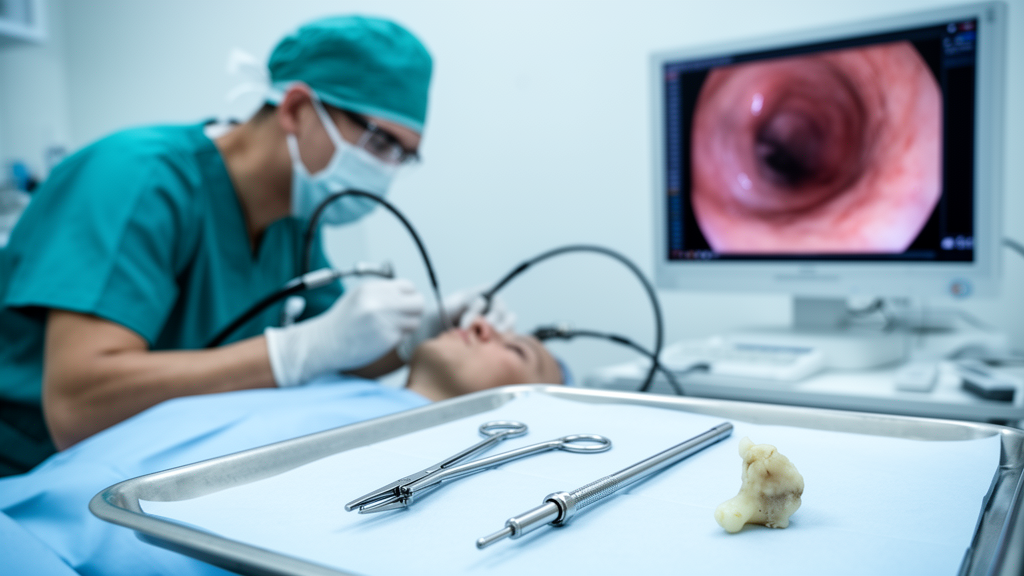
After the patient signed an informed consent form, the broncholith was removed via flexible bronchoscopy under local anaesthesia. Bronchoscopic examination showed the stone surrounded by granulation tissue and firmly anchored at its base. Because the broncholith was tightly adherent to the bronchial wall and the surrounding granulation tissue, and because it lay near several branches of the pulmonary vessels, the team judged that direct removal of the entire stone in one piece would increase the risk of bleeding.
The technical core of the procedure was the use of a 1.1 mm diameter disposable cryoprobe, operated with carbon dioxide, applied directly to the exposed broncholith. The probe was first applied for 3 to 5 seconds. When the stone proved difficult to fragment, the freezing time was extended to 5 to 10 seconds.
The cycle of cryoprobe-assisted fragmentation followed by forceps extraction was repeated 2 to 4 times, allowing most of the broncholith to be removed in pieces without forceful pulling. To minimize the risk of major bleeding, small residual fragments at the base were deliberately left in place rather than forcibly extracted. Only minor bleeding was observed throughout the entire procedure.
The outcome was encouraging on both immediate and follow-up measures. Subsequent haematologic evaluation confirmed a diagnosis of immune thrombocytopenia. A follow-up chest CT scan obtained one month after the procedure showed that the calcified lesion had disappeared, and the patient had not experienced any further episodes of haemoptysis.
Bronchoscopic images taken after the removal showed no visible residual broncholith, and the extracted fragments confirmed the piecemeal nature of the extraction. For a patient whose preoperative platelet count sat below 50 × 10^9/L, the absence of significant bleeding was the clinically decisive result. The discussion accompanying the case places the technique in context.
Several methods have been described for broncholith extraction, including biopsy forceps, balloon catheters, laser treatment and cryotherapy. Previous studies have shown that cryotherapy may be useful for broncholiths that are fixed or partially attached and difficult to remove with forceps alone. By helping to separate or fragment the stone, cryotherapy can avoid forcible traction when there is a high risk of bleeding.
The authors note that their case further suggests this approach may be feasible in certain patients at high risk of bleeding, such as those with thrombocytopenia, although a single case cannot establish a safe platelet threshold for the procedure. Choice of scope was itself a deliberate decision. Rigid bronchoscopy has been reported to achieve a higher success rate than flexible bronchoscopy for broncholith removal, but the stone in this patient sat in a distal bronchus.
The team therefore selected flexible bronchoscopy using a cryoprobe combined with biopsy forceps. The authors emphasize that careful preparation was central to the plan: performing contrast-enhanced CT with reconstruction before the procedure, conducting a multidisciplinary assessment, and preparing bronchial occlusion balloons in case major bleeding developed. Intraoperatively, they recommend that cryotherapy target the exposed broncholith, that freezing time be adjusted according to how difficult fragmentation proves, and that strong traction be avoided throughout.
The distal reach of the technique is one of its more notable features. Previous reports have described cryoprobe-assisted broncholith removal in the right middle lobe bronchus, the right upper lobe bronchus and the left upper lobe lingular segment bronchus. In this case, the procedure was performed in fifth to seventh generation bronchi, the progressively finer subdivisions of the airway tree, suggesting that the approach may also be feasible in more distal bronchi than those covered by earlier reports.
The main advantage of cryoprobes, the authors argue, lies in their ability to facilitate broncholith fragmentation by freezing, allowing larger stones to be removed piecemeal and reducing the need for forceful traction and the bleeding risk that comes with it. This may offer an alternative to forceful en bloc extraction for firmly attached broncholiths or for patients whose bleeding risk is elevated. The authors are careful about the limits of what a single case can show.
The report cannot define a safe platelet threshold for broncholith removal, and the successful outcome in one patient with a count of 49 × 10^9/L does not establish that such counts are safe for all patients or all procedures. What the case does suggest is that broncholith removal under flexible bronchoscopy using a cryoprobe combined with biopsy forceps may be feasible in selected patients with thrombocytopenia, provided that preoperative imaging, multidisciplinary planning and bleeding preparations are in place. The authors also note that if the procedure proves unsuitable under local anaesthesia, rigid bronchoscopy should be considered as an alternative.
For now, the case stands as a documented example of a piecemeal, traction-free extraction in a high-risk patient, and as a data point for clinicians weighing how far to push interventional bronchoscopy in the thrombocytopenic airway.
Subject of Research: Bronchoscopic removal of a broncholith using a cryoprobe combined with biopsy forceps in a patient with immune thrombocytopenia
Article Title: Broncholith Removal Using a Cryoprobe in Combination With Biopsy Forceps in a Patient With Immune Thrombocytopenia: A Case Report
Article References: Xiao, Q., Ma, N., & Zhu, J. (2026). Broncholith Removal Using a Cryoprobe in Combination With Biopsy Forceps in a Patient With Immune Thrombocytopenia: A Case Report. Respirology Case Reports, 14(10), Article e70779. https://doi.org/10.1002/rcr2.70779
Image Credits: AI Generated
DOI: 10.1002/rcr2.70779
Keywords: broncholithiasis, bronchoscopy, cryoprobe, cryotherapy, immune thrombocytopenia, haemoptysis, pulmonary tuberculosis, interventional pulmonology, case report, airway obstruction, chest CT, bleeding risk
News Source: Ophelia Keating. (October 25, 2026). Research report: Broncholith Removal Using a Cryoprobe in Combination With Biopsy Forceps in a Patient With Immune Thrombocytopenia: A Case Report. Scienmag.