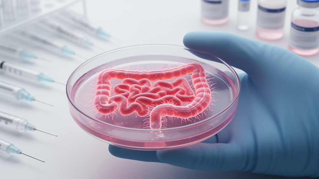
For decades, the study of intestinal infections has been constrained by a stubborn mismatch: the laboratory models used to investigate enteric pathogens rarely behave like the human gut they are meant to represent. A new review by Linus Schweizer, Ashley Gilliland, Yan Chen, Artemis Melville-Bowser and Bruce Vallance of the University of British Columbia, published in PLOS Pathogens, argues that two bioengineered platforms, air-liquid interface (ALI) cultures and gut-on-a-chip (GoC) microfluidic systems, are now closing that gap. The authors collectively term these systems next-generation in vitro models, or NGIMs, and show how they recreate critical features of the human gastrointestinal mucosal surface, including a thick mucus layer, crypt-villus architecture and the biomechanical forces that shape infection. Their application to bacterial, fungal, viral and parasitic pathogens has already revealed mechanisms that traditional cell lines and rodent models failed to detect.
The limitations of conventional models are well documented. Widely used intestinal epithelial cell lines such as Caco-2 secrete little if any mucus, while HT29-MTX cells produce mucins characteristic of the gastric and respiratory mucosa rather than the intestine. Host tropism presents another barrier: human norovirus fails to establish infection in mice, and enterotoxigenic Escherichia coli (ETEC) infection in mice does not reproduce the characteristic diarrheal symptoms seen in humans. Animal models also show significant variability between laboratories, often driven by differences in gut microbiota composition. Intestinal organoids derived from patient tissue are a promising alternative, particularly as the FDA Modernization Act 2.0 and the National Institutes of Health push toward reducing animal use, but they are difficult to infect because their apical surface and mucus face inward, requiring laborious micro-injection into individual organoid lumens, and they exhibit considerable heterogeneity in size and cellular composition even within a single experiment.
ALI cultures solve the orientation problem by growing intestinal epithelial monolayers on Transwell inserts, supplied with media basolaterally while the apical side is exposed to air. This drives the formation of crypt-like structures, rapid differentiation into goblet cells, mucin release and the development of an overlying mucus layer within 14 to 28 days. GoC systems take a different route: organoid fragments are seeded into the upper channel of a porous membrane with endothelial cells below, and continuous fluid flow and stretch forces promote epithelial maturation by day five. Both platforms yield a well-differentiated epithelium with correct apical-basal polarity, cellular heterogeneity and a robust mucus layer, and both can be adapted to support an apical anaerobic compartment, enabling the study of obligate anaerobic pathogens and commensal microbiota that cannot be cultured under traditional aerobic conditions.
The importance of gut architecture has become strikingly clear in these systems. Infections by Clostridioides difficile and Listeria monocytogenes decrease stem cell-mediated regeneration at the crypt base, reducing pathogen clearance from this niche and potentially increasing susceptibility to secondary infections. Listeria expresses virulence factors to specifically invade villus tips, while Cryptosporidium induces microvilli elongation that may support parasitic proliferation by enhancing nutrient uptake. Traditional cell culture forms only flat monolayers lacking these structures. In GoC systems, the three-dimensional villi-like architecture proved necessary for Shigella flexneri invasion during early infection, and villi were also critical for coxsackievirus B1, which preferentially shed from infected cells at the apex of villi. By recapitulating the crypt-villus axis, NGIMs capture niche-specific pathogen behavior that flat cultures cannot.
Cellular differentiation is equally decisive. Many enteric pathogens target specific epithelial cell subsets, an aspect easily overlooked in undifferentiated cultures. Human BEST4/CA7+ cells, recently identified as targets for heat-stable enterotoxin STa produced by ETEC, are absent in mice, yet NGIMs can promote differentiation of stem cells into all intestinal epithelial lineages and recapitulate region-specific populations along the small to large intestinal axis. Compared with three-dimensional organoids, ALI and GoC cultures display a more mature phenotype, with reduced proliferative signatures and greater transcription of nutrient transport and metabolism genes. The mucus barrier tells a similar story of species specificity: the EatA serine protease encoded by ETEC degrades human MUC2 but fails to degrade mouse Muc2 because of structural differences, and the thick colonic mucus of humans is comparably thinner in mice even accounting for size. GoC systems using human colonic organoids can even form a mucus bilayer mimicking the penetrable outer and impenetrable inner layers of the native colon.
These physiological features have exposed previously hidden infection strategies. In ALI cultures, exposure to mucus promotes C. difficile persistence by shifting its metabolism toward mucus-derived nutrients while stimulating biofilm formation and altering mucus viscoelasticity. A GoC model of stable co-colonization with the anaerobe Bacteroides fragilis revealed that vancomycin treatment drove C. difficile into dense biofilms clustered with vancomycin-resistant B. fragilis, and some C. difficile invaded epithelial cells to become intracellular, illustrating a potential mechanism of antibiotic evasion and infection recurrence. GoC studies also showed that S. flexneri microcolonies start from a single bacterium and invade cells via direct intracellular basal translocation rather than the paracellular route previously assumed, and that the pathogen exploits human antimicrobial peptide HD5-induced filopodial extension for invasion. Exposing Citrobacter rodentium to ALI-derived mucus identified a previously unrecognized mucolytic function for the serine protease EspC.
Parasites and viruses have yielded equally notable discoveries. Mouse-derived ALI monolayers supported complete development of Cryptosporidium parvum, which expanded 100-fold and generated viable oocysts, revealing a previously unidentified life stage termed late-stage macrogamonts and enabling in vitro genetic crossing without animal hosts. GoC systems permit long-term culturing of both C. parvum and the human-specific Cryptosporidium hominis for periods ranging from 10 days to 4 weeks. Organoid-derived Transwell cultures captured Trichuris muris larvae degrading the overlying mucus, invading epithelial cells and forming syncytial tunnels characteristic of in vivo infection, while live imaging in microfluidic devices showed larvae tunneling toward the base of crypts and causing localized cell death. On the viral front, porcine ALI monolayers revealed that susceptibility to transmissible gastroenteritis virus and porcine epidemic diarrhea virus was reduced by the antiviral effects of sialylated mucus, and human iPSC-derived ALI monolayers resembling fetal-like tissue expressed less ACE2, the SARS-CoV-2 receptor, resulting in lower infection rates consistent with milder COVID-19 in children. Mouse-derived ALI cultures supported murine norovirus infection, revealing tuft cell tropism and a reservoir mechanism for persistent infection.
Biomechanical forces and commensal microbes add further layers of realism. In GoC models with peristalsis-like stretch, commensal E. coli persistently colonized the lumen without harming barrier function, whereas enteroinvasive E. coli multiplied dramatically at the epithelial surface, destroying villi. Under flow conditions, S. flexneri increased intracellular propagation and Type 3 secretion system expression, Campylobacter jejuni induced metabolic reprogramming of host cells absent in static infections, and fluid flow enhanced the potency of ETEC STa on more polarized cells. Commensal strains also modulate pathogens: Lactobacillus rhamnosus GG co-aggregated with enterotoxigenic Bacteroides fragilis and impaired its pathogenicity, pre-colonization with L. rhamnosus suppressed Candida albicans yeast-to-hyphal transition, and metabolites from human, but not murine, microbiota induced enterohemorrhagic E. coli chemotaxis and motility genes in human GoC models. The authors caution, however, that microbial communities in NGIMs remain less complex and less stable than the native intestinal microbiome, and that mechanical stretch itself can compromise barrier integrity, warranting careful controls.
Choosing between the two platforms depends on the question at hand. ALI cultures excel for mucus studies because the layer is readily accessible for in situ rheology, and they are cost-effective, relying on standard Transwell equipment and offering high-throughput scalability. GoC systems uniquely capture peristalsis and luminal flow, key contributors to pathogen clearance, but require greater technical expertise, dedicated pumps and flow controllers, and significant financial investment. Both systems currently lack a fully functional integrated immune component, interactions with distal organs and a stable complex microbiome, so the review positions NGIMs as complementary to, rather than complete replacements for, in vivo models. Looking ahead, the authors highlight the integration of stromal and tissue-resident immune cells, which recently recapitulated celiac disease enteropathy in duodenal ALI cultures, the enteric nervous system, whose disruption by C. difficile toxins reduces gut mobility and aids colonization, real-time TEER, pH and oxygen sensors, genetic reporter lines, and multi-organ microfluidic systems that have already modeled Shiga toxin-induced hemolytic-uremic syndrome. Cheaper chip fabrication via 3D bioprinting and standardized protocols for media and extracellular matrix substrates should improve accessibility and inter-laboratory reproducibility.
The precision medicine potential is tangible. ALI monolayers grown from the colonic biopsies of ulcerative colitis patients mimicked disease phenotypes in vitro, including enhanced susceptibility to an E. coli pathobiont, and patient-specific differences in mucus development influenced infection outcomes. The authors envision long-term co-culture of patient-derived epithelium with an autologous microbiome to guide antimicrobial regimens that minimize microbiome disruption, reducing the risk of post-antibiotic infections such as C. difficile. The physiological mucus layer also makes NGIMs well suited for evaluating bacteriophage therapy, antimicrobial peptides with enhanced activity in the presence of mucus, anti-adhesin therapies and microbiota-modulating interventions including probiotics and fecal microbiota transplantation. By bridging the gap between conventional methods and the complexities of the human gut, ALI and GoC systems are providing new insights into both the virulence and the weaknesses of enteric pathogens that pose a global threat to human health.
Subject of Research: Next-generation human in vitro models for studying enteric pathogen infections
Article Title: Rebuilding the gut: Decoding enteric pathogenesis using next-generation human in vitro models
Article References: Schweizer, L. H., Gilliland, A., Chen, Y., Melville-Bowser, A., & Vallance, B. A. (2026). Rebuilding the gut: Decoding enteric pathogenesis using next-generation human in vitro models. PLOS Pathogens, 22(10), e1014718. https://doi.org/10.1371/journal.ppat.1014718
Image Credits: AI Generated
DOI: 10.1371/journal.ppat.1014718
Keywords: gut-on-a-chip, air-liquid interface, enteric pathogens, intestinal organoids, mucus barrier, infection biology, microfluidics, antimicrobial resistance, pathogen-commensal interactions, precision medicine, Rebuilding, Decoding
News Source: Kristina Jarvis. (October 26, 2026). Rebuilding the gut in miniature: next-generation models decode enteric pathogenesis. Scienmag.