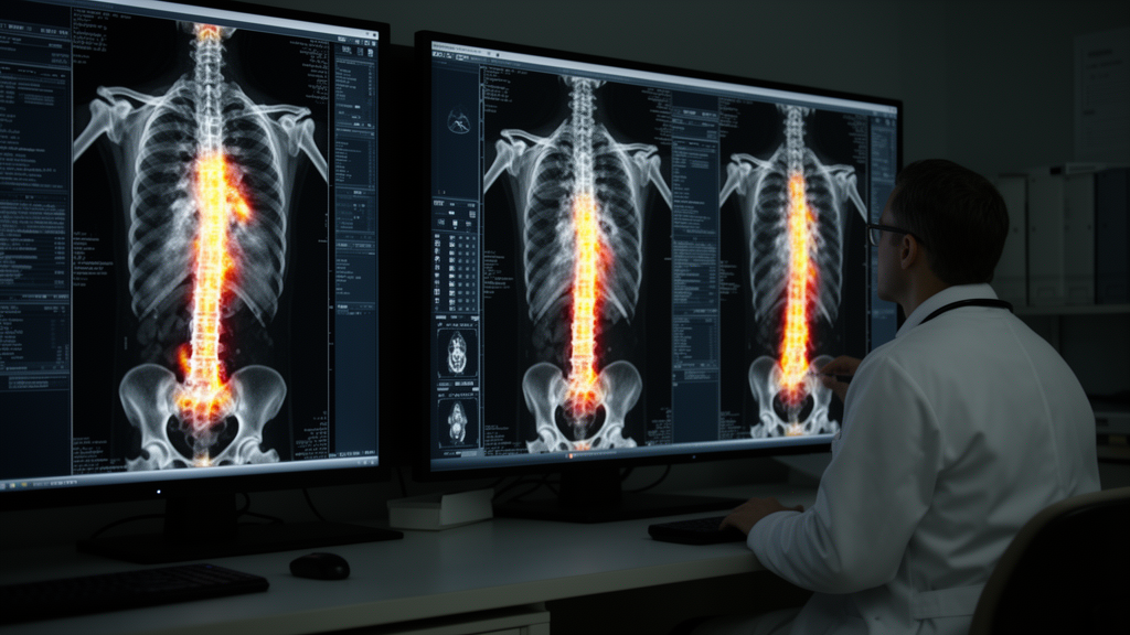
A [18F]FDG-PET/CT scan performed in a 64-year-old woman being evaluated for fever of unknown origin revealed extensive osteosclerosis of both the axial and appendicular skeleton, with markedly increased skeletal metabolic activity reaching an SUVmax of 17.5. The imaging also showed several punched-out osteolytic lesions in the skull and ribs, some of which had a soft tissue component. The case, published as an image article in EJNMMI Reports, documents an unusually striking skeletal presentation of an exceptionally rare leukemia.
The clinical picture that prompted the scan included severe thrombocytopenia, with a platelet count of 15 × 10⁹/L, a normal white blood cell count with a left shift, 2% blasts on manual differential of the peripheral blood, and markedly elevated lactate dehydrogenase exceeding 50 µkat/L. Initial sternal bone marrow aspiration was non-diagnostic. A subsequent iliac bone marrow aspiration identified myeloid blasts with an immunophenotype of CD41+/CD61+/CD56+/CD81+/CD117+, consistent with a diagnosis of acute megakaryoblastic leukemia (AMKL).
AMKL is described in the report as an extremely rare and aggressive subtype of acute myeloid leukemia, occurring predominantly in children with Down syndrome. The authors explain that cytokines derived from megakaryoblasts, particularly transforming growth factor-β and platelet-derived growth factor, promote bone marrow fibrosis and osteosclerosis, providing a biological link between this leukemia subtype and the dense bone appearance seen on the scan.
The authors note that patients with myeloid malignancies are infrequently evaluated with [18F]FDG-PET/CT. Potential indications include extramedullary infiltration of acute myeloid leukemia, systemic mastocytosis, and extramedullary hematopoiesis in myeloproliferative neoplasms. Related tracers have also shown prognostic value elsewhere in myeloid disease: [18F]FLT-PET/CT has demonstrated prognostic value in primary myelofibrosis.
Diffuse osteosclerosis on imaging carries a broad differential diagnosis, including skeletal metastases, endocrine causes such as hyperparathyroidism, renal osteodystrophy, fluorosis, and Paget disease. Although [18F]FDG-PET/CT is not routinely performed in patients with myeloid malignancies, the authors argue that recognizing diffuse osteosclerosis with increased skeletal metabolic activity may raise suspicion of osteosclerosis-associated hematological disorders and prompt timely histopathological evaluation. To their knowledge, diffuse osteosclerosis with intense skeletal [18F]FDG uptake has been only rarely reported in adults with AMKL, making this adult case a notable addition to the limited published experience.
Subject of Research: An imaging case report of diffuse osteosclerosis on FDG-PET/CT in an adult with acute megakaryoblastic leukemia.
Article Title: Diffuse osteosclerosis in acute megakaryoblastic leukemia on [18 F]FDG-PET/CT
Article References: Horňák, G., Ičová, V., Burianková, E., Látal, V., Szotkowská, R., & Kamínek, M. (2026). Diffuse osteosclerosis in acute megakaryoblastic leukemia on [18 F]FDG-PET/CT. EJNMMI Reports, 10(1), Article 45. https://doi.org/10.1186/s41824-026-00333-4
Image Credits: AI Generated
DOI: 10.1186/s41824-026-00333-4
Keywords: FDG-PET/CT, acute megakaryoblastic leukemia, osteosclerosis, acute myeloid leukemia, bone marrow fibrosis, osteolytic lesions, thrombocytopenia, myeloid malignancies, nuclear medicine, case report, Diffuse, acute
News Source: Nathaniel Bowman. (October 27, 2026). Rare leukemia revealed by diffuse osteosclerosis on FDG-PET/CT. Scienmag.