In a laboratory study published in Immunity, Inflammation and Disease, researchers report that Sini Decoction, a classical Chinese herbal formula, changed the inflammatory behavior of macrophages in a cell model of atherosclerosis and appeared to act through the transcriptional corepressor NCOR1 and mitochondria-related pathways. The work, conducted entirely in cultured RAW 264.7 macrophage cells, offers a molecular starting point for understanding how the traditional formula might influence vascular inflammation, while its authors are explicit that the findings cannot yet be extended to plaques in living blood vessels.
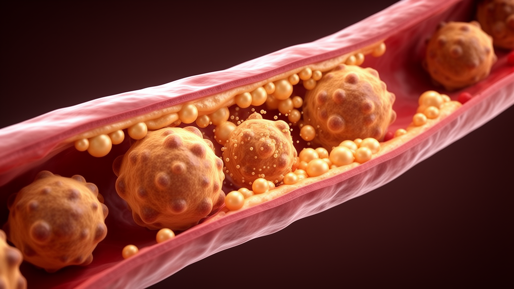
Lower extremity atherosclerosis, a major subtype of peripheral artery disease, involves progressive arterial stenosis, vascular sclerosis, and thrombotic occlusion in the leg vasculature, producing insufficient blood flow and symptoms ranging from intermittent claudication and muscle weakness to severe ischemic ulcers and necrosis. The condition visibly affects quality of life and acts as a risk amplifier for systemic cardiovascular morbidity. Pathologically, lipid deposits accumulate in the arterial intima, smooth muscle cells proliferate, fibrous tissue forms, and immune cells including macrophages infiltrate the vessel wall, ultimately forming plaques that can narrow the lumen and, in severe cases, trigger thrombosis and tissue ischemia or necrosis.
Macrophages occupy a central position in this disease process. These immune cells play different roles at different stages of atherosclerosis, particularly in plaque formation, stabilization, and rupture, and their polarization state visibly affects plaque stability. M1-polarized macrophages are considered the main pro-inflammatory cell type in atherosclerosis, exacerbating local inflammatory responses through the secretion of large amounts of inflammatory cytokines. Current pharmacological management of lower extremity atherosclerosis relies heavily on statins and antiplatelet drugs, but existing regimens often fail to completely reverse plaque formation and stabilization and may be accompanied by side effects, motivating the search for new targets that stabilize plaques by regulating immune and inflammatory responses.
Sini Decoction originates from the Treatise on Febrile Diseases and is known for warming, blood-activating, and circulation-improving effects, having been used historically for cold syndromes caused by yang deficiency such as abdominal pain due to cold-dampness. In recent years it has been widely used as an adjunct therapy for various cardiovascular diseases, and prior studies suggest it can slow atherosclerosis progression through multiple pathways, including regulating endothelial cell function, inhibiting macrophage activation, and reducing blood lipids. However, the specific mechanisms by which the formula regulates macrophage polarization and plaque stability remained unclear, which is the gap the new study set out to address.
The researchers focused on nuclear receptor corepressor 1, or NCOR1, a key transcriptional corepressor implicated in macrophage inflammatory phenotype switching and mitochondrial homeostasis. Previous work cited in the paper found that macrophage-specific NCOR1 deficiency significantly exacerbated atherosclerotic lesions in mice, with increased foam cell formation, upregulated pro-inflammatory cytokine expression, enlarged necrotic cores, and thinner fibrous caps; mechanistically, NCOR1 exerts protective effects mainly by inhibiting the pro-atherogenic activity of peroxisome proliferator-activated receptor gamma. NCOR1 expression is also decreased in human atherosclerotic plaques, with the reduction more pronounced in ruptured plaques than in non-ruptured ones, and a complementary study linked NCOR1 to glucose metabolic reprogramming in macrophages.
Methodologically, the team established an inflammatory model by stimulating RAW 264.7 macrophages with 1 microgram per milliliter of lipopolysaccharide for 24 hours. Cells were then randomly assigned to a blank group, a model group, and low-, medium-, and high-dose Sini Decoction-medicated serum groups, with three independent biological replicates per group. The medicated serum was prepared by decocting the crude herbal materials twice with water, concentrating and lyophilizing the filtrates, administering the decoction to healthy rats by gavage while controls received saline, and then collecting, inactivating, filtering, and storing the serum. After LPS stimulation, treated groups received the medicated serum for an additional 24 hours while blank and model groups received equal volumes of blank rat serum.
The results showed a coherent dose-dependent pattern. Compared with the blank group, LPS-stimulated model cells displayed significantly increased mRNA expression of the pro-inflammatory cytokines IL-6 and TNF-α (p = 0.002 and p = 0.003) and of the M1 macrophage markers CD16/32 and COX2 (p = 0.018 and p < 0.001), alongside significantly decreased IL-10 (p = 0.001) and the M2 markers Arg1 and CD206 (p = 0.001 and p = 0.002). With increasing doses of the medicated serum, IL-6 and TNF-α progressively fell, IL-10 rose, M1 markers declined, and M2 markers climbed, with all dose groups differing significantly from the model group and the high-dose group no longer differing from the blank group on any parameter.
Mitochondrial measurements pointed in the same direction. In the model group, protein levels of the mitophagy-related proteins LC3, Parkin, and FUNDC1 were significantly elevated (p = 0.002, p < 0.001, and p = 0.003) while P62 was significantly reduced (p = 0.004); Sini Decoction-medicated serum reversed each of these changes dose-dependently. Seahorse mitochondrial stress testing revealed that the model group had significantly decreased oxygen consumption rate and significantly increased extracellular acidification rate (both p < 0.001), and the serum intervention restored OCR while lowering ECAR across doses. Cell viability, measured by MTS assay, was significantly reduced by LPS (p < 0.001) and recovered progressively with treatment. NCOR1 followed the same trajectory: mRNA fell from 1.00 ± 0.08 in blank cells to 0.36 ± 0.05 in the model group and recovered to 0.95 ± 0.09 at the high dose, with protein gray-value ratios moving from 0.97 ± 0.07 to 0.31 ± 0.04 and back to 0.92 ± 0.08.
The authors interpret these findings as evidence that Sini Decoction-containing serum dose-dependently inhibited the pro-inflammatory response of macrophages, promoted polarization toward the M2 anti-inflammatory phenotype, ameliorated abnormal mitophagy and mitochondrial function, and upregulated NCOR1 expression, suggesting the formula’s regulatory effects may be related to the NCOR1 pathway. They caution, however, that the study examined only static protein expression without mitophagic flux monitoring, so the upregulation of P62 cannot be directly interpreted as enhanced mitophagy and may instead indicate a blockade of the autophagic degradation pathway for damaged mitochondria. Because no NCOR1 gene intervention experiments were performed, it is not yet possible to confirm that the formula acts by upregulating NCOR1 to regulate mitophagy.
The limitations are substantial and acknowledged in detail. The work relied solely on RAW 264.7 macrophages in vitro, without an animal model of lower extremity atherosclerosis, and a single-cell culture environment cannot fully recapitulate the complex physiological microenvironment of human blood vessels. No clinical specimens from patients were analyzed, medicated serum was used without isolating the active monomers, so the core pharmacologically active substances were not identified, and the concentration gradient was coarse, making the optimal dose difficult to pinpoint. The authors state that future work will establish an ApoE-deficient mouse atherosclerosis model, purify the formula’s characteristic active components, add NCOR1 gene intervention and rescue experiments, use mt-Keima fluorescent probes and bafilomycin A1 blockade assays to measure mitophagic flux, and collect clinical vascular specimens. For now, the study stands as in vitro molecular evidence that a centuries-old formula can shift macrophage behavior in a dish, with any claim about stabilizing plaques in human arteries still awaiting in vivo and clinical verification.
Subject of Research: Effects of Sini Decoction-containing serum on macrophage polarization, mitophagy-related proteins, and NCOR1 expression in an LPS-induced RAW 264.7 cell model of atherosclerotic inflammation
Article Title: Mechanism of Si Ni Decoction in Stabilizing Atherosclerotic Plaques in the Lower Extremities by Regulating Macrophage Polarization via NCOR1 Mitophagy
Article References: Liu, Z., Ge, S., Yang, J., Zhou, Y., Jin, L., & Fang, Y. (2026). Mechanism of Si Ni Decoction in Stabilizing Atherosclerotic Plaques in the Lower Extremities by Regulating Macrophage Polarization via NCOR1 Mitophagy. Immunity, Inflammation and Disease, 14(10), Article e70546. https://doi.org/10.1002/iid3.70546
Image Credits: AI Generated
DOI: 10.1002/iid3.70546
Keywords: Sini Decoction, atherosclerosis, macrophage polarization, NCOR1, mitophagy, RAW 264.7 macrophages, peripheral artery disease, lower extremity atherosclerosis, mitochondrial function, traditional Chinese medicine, inflammation, in vitro study
News Source: Ophelia Keating. (October 27, 2026). Ancient Chinese Formula Sini Decoction Rewires Immune Cells in Lab Study of Atherosclerosis. Scienmag.