Quercetin treatment was associated with reduced acute inflammatory and extracellular matrix-remodeling responses 24 hours after collagenase-induced intracerebral hemorrhage (ICH) in mice, according to a study combining transcriptomics-informed network pharmacology, experimental validation and assessment of human ICH samples. The work was preclinical: it did not test quercetin as a treatment in patients with ICH.
Researchers first integrated 4,646 predicted quercetin targets with 1,602 ICH-associated genes, identifying 657 shared genes that mapped to 642 mouse orthologs. They combined these targets with exploratory differential-expression results from one mouse brain transcriptomic dataset and an endoplasmic-reticulum-stress-related reference set, producing 29 candidate genes. Functional enrichment implicated TNF and IL-17 signaling, leukocyte migration, stress responses and extracellular matrix processes.
A protein-protein interaction analysis ranked the 29 candidates using five centrality methods. Seven genes appeared in all five top-10 lists and were designated consensus hubs: Il1b, Mmp9, Timp1, Myc, Fos, Hmox1 and Socs3. In the discovery dataset, five hubs had higher expression after ICH than in controls and each had an area under the ROC curve greater than 0.9. In an independent dataset, six hubs were differentially expressed and also had AUC values above 0.9, although these exploratory analyses involved only five ICH and five control samples per dataset.
Molecular docking produced putative binding poses between quercetin and all seven hub proteins. The strongest predicted binding scores were reported for Fos, Hmox1, Il1b, Mmp9 and Timp1, while Socs3 and Myc had moderate predicted affinity. These computational estimates were interpreted as hypothesis-generating and do not demonstrate direct biochemical binding or target engagement.
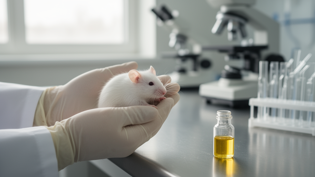
For experimental validation, researchers used male C57BL/6 mice aged 6 to 8 weeks. ICH was induced by injecting collagenase into the striatum, with sham-operated animals receiving saline. Groups of six mice received intraperitoneal quercetin at 25, 50 or 100 mg/kg per administration, twice at 0.5 and 12 hours after hemorrhage, for total 24-hour administered doses of 50, 100 and 200 mg/kg. A separate group received dexamethasone at 2 mg/kg per administration on the same schedule as an anti-inflammatory comparator. The quercetin and dexamethasone regimens were described as literature-informed exploratory designs rather than direct replications.
At 24 hours, brain water content was significantly higher after ICH than in sham-operated mice. Quercetin at 100 mg/kg per administration reduced brain water content compared with untreated ICH, as did dexamethasone; the difference between those two groups was not significant. In perihematomal tissue, quercetin at 50 and 100 mg/kg reduced Il1b expression, while all three doses reduced Mmp9 and Fos expression. Timp1 expression increased after all three doses. Hmox1 increased selectively at the high dose, and Socs3 rose dose-dependently, while Myc was not significantly altered by treatment.
Serum measurements in mice showed increases in IL-1β, IL-6, TNF-α and MMP-9 after ICH. The 25 mg/kg quercetin dose did not significantly reduce any of these markers relative to untreated ICH. At 50 mg/kg, quercetin reduced all four markers, while at 100 mg/kg it produced reductions in all four that were not significantly different from sham levels. Dexamethasone showed a similar pattern, but the study did not establish equivalence between the treatments.
To assess disease relevance in humans, investigators analyzed paired perihematomal and remote non-hemorrhagic brain tissues from six ICH patients undergoing clinically indicated hematoma evacuation. Perihematomal tissue had higher IL1B, MMP9, TIMP1 and HMOX1 messenger RNA than matched remote tissue. Western blotting confirmed higher IL-1β, MMP-9 and TIMP1 proteins, and immunohistochemistry showed greater MMP-9 immunoreactivity and a larger MMP-9-positive area in perihematomal samples. These observations support the relevance of selected targets to human ICH pathology, but the patients did not receive quercetin, so the tissue data do not demonstrate quercetin-specific efficacy or target engagement.
Serum was also collected within 24 hours of symptom onset from six ICH patients and six age- and sex-approximated healthy controls. IL-1β, IL-6, IL-10, IL-17A and TNF-α concentrations were higher in patients. The human cohort was small and observational, while the mouse experiments were limited to males, one collagenase model, small groups of six and a single 24-hour endpoint. Hematoma or lesion volume, neurological and behavioral outcomes, histopathology, blood-brain barrier integrity and cell-specific mechanisms were not assessed. The results therefore support an acute experimental association between quercetin and lower inflammatory, matrix-remodeling and edema-related markers, not clinical benefit in people with ICH.
Subject of Research: Preclinical assessment of quercetin in experimental intracerebral hemorrhage
Article Title: Quercetin attenuates acute inflammation and extracellular matrix remodeling after intracerebral hemorrhage: transcriptomics-informed network pharmacology and experimental validation
Article References: Shu, J., Feng, K., Pan, A., Li, G., Liu, K., & Feng, L. (2026). Quercetin attenuates acute inflammation and extracellular matrix remodeling after intracerebral hemorrhage: transcriptomics-informed network pharmacology and experimental validation. Frontiers in Pharmacology, 17, Article 1930434. https://doi.org/10.3389/fphar.2026.1930434
Image Credits: AI Generated
DOI: 10.3389/fphar.2026.1930434
Keywords: intracerebral hemorrhage, quercetin, neuroinflammation, extracellular matrix, network pharmacology, mouse model, MMP-9, IL-1β, human brain tissue, transcriptomics, attenuates, acute
News Source: Cassandra Pierce. (October 21, 2026). Quercetin reduces acute inflammatory and matrix-remodeling markers in mouse intracerebral hemorrhage. Scienmag.