


Gadolinium, the metal at the heart of many magnetic resonance imaging scans, can now be tracked at remarkably low concentrations thanks to a new sensor developed by researchers at the German Cancer Research Center (DKFZ) in Heidelberg. The team, led by Leif Schröder, head of the Department of Translational Molecular Imaging, reports that its approach detects gadolinium in the micromolar range under experimental conditions, a sensitivity the researchers say surpasses what was previously achievable with MRI-based detection methods.
The finding matters because gadolinium-based contrast agents are used in just under half of all MRI scans. The element provides the contrast that makes tumors and other pathological changes clearly distinguishable from healthy tissue. Enclosed within a chelate molecule, a kind of molecular cage, gadolinium interacts through its magnetic properties with water molecules inside the scanner, causing tissues with high water content to appear brighter in the resulting images.
After the examination, the contrast agents are excreted via the kidneys in the urine and end up in wastewater. There, the gadolinium remains bound within its chelate cage over the long term. Although the element stays caged, the DKFZ team argues that sensitive analytical tools are needed to follow its path through the environment. “Nevertheless, we see an urgent need to lay the foundations for new and sensitive analytical methods that will enable us to track gadolinium in the environment – not only from contrast agents but also from industrial processes,” Schröder explains.
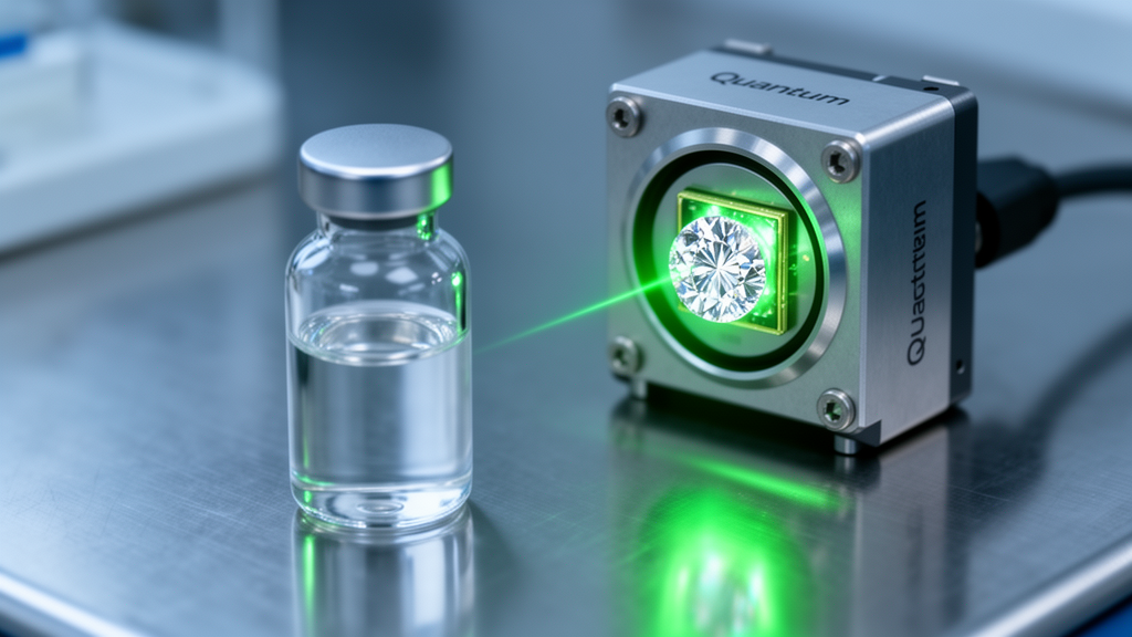
The new sensor relies on an unusual pairing of physics and chemistry. The researchers used hyperpolarized xenon, a noble gas that has been strongly magnetized and is itself enclosed within a different type of cage molecule that produces signals in MRI. Inside this setup, the nuclear spin of the xenon acts as a quantum sensor, yielding two distinct signals: one from xenon bound inside the cage molecules and one from free xenon in the surrounding solution.
Gadolinium then enters the picture as a competitor. The metal displaces xenon from its cage, and as the displacement proceeds, the corresponding xenon signal diminishes and eventually disappears altogether. By monitoring this decline, the researchers can work backwards to the amount of gadolinium present in the sample.
“From the reduction in the xenon signal, we were able to determine the concentration of gadolinium outside the cage,” says Hannah Gerbeth, lead author of the study. The team found that gadolinium concentrations in the micromolar range can be measured this way, making the cage molecule a highly sensitive gadolinium sensor under the conditions of the experiment. “This discovery shows that the unique properties of the interaction between gadolinium, xenon and the cage enable more sensitive detection than was possible in previous research using MRI,” Schröder adds.
The technique is described in a competition assay format using a cage molecule known as cryptophane-A monoacid, and the detection was carried out with HyperCEST nuclear magnetic resonance spectroscopy. The work was published in the journal Sensors & Diagnostics as a peer-reviewed experimental study, with the article appearing on 30 July 2026.
Beyond environmental monitoring, the researchers see potential clinical relevance. The method can distinguish free gadolinium from chelated forms, which could help improve both the clinical application of contrast agents and the detection of gadolinium-containing substances in the environment.
The work remains at an early stage, and the reported sensitivity applies to the conditions of the laboratory experiment rather than to field measurements. The DKFZ team plans to test whether the sensor works across other concentration ranges, and it also intends to investigate whether other ions are selectively bound by the cage and could therefore interfere with gadolinium detection.
The study was conducted at the German Cancer Research Center, Germany’s largest biomedical research institute with more than 3,500 employees, which is funded primarily by the Federal Ministry of Research, Technology and Space and by the state of Baden-Württemberg. The researchers report no conflicts of interest.
Subject of Research: A hyperpolarized xenon-based cage sensor for detecting gadolinium from MRI contrast agents in the micromolar range
Article Title: Highly sensitive detection of MRI contrast agents
Article References: Highly sensitive detection of MRI contrast agents. Original publication (Bibliographic details unavailable.)
Image Credits: AI Generated
DOI: Not provided
Keywords: gadolinium, MRI contrast agents, hyperpolarized xenon, quantum sensor, HyperCEST NMR, cryptophane, environmental detection, wastewater, German Cancer Research Center, chelate, molecular imaging, Sensors & Diagnostics
News Source: Katie Riggs. (October 30, 2026). Quantum Sensor Spies Traces of MRI Contrast Agent Gadolinium in Water. Scienmag.



