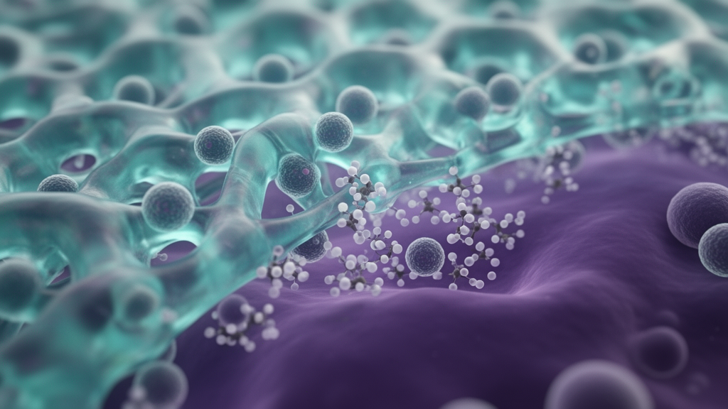
In an extensive narrative review published in Frontiers in Biomaterials Science, researcher Moawiah M. Naffaa outlines an integrative framework for programmable immune biointerfaces, shifting the foundational concept of biocompatibility from passive host tissue tolerance toward the precise instruction of immune-cell state trajectories over space and time. Traditional evaluations in biomaterials science have historically relied upon the absence of overt cytotoxicity, foreign body encapsulation, acute inflammatory destruction, or systemic rejection-like responses. However, modern functional outcomes reveal that materials once classified as chemically inert can still promote chronic low-grade inflammation, persistent fibrotic walling-off, impaired vascularization, and structural isolation. To overcome these limitations, the review defines a programmable immune biointerface as an engineered biomaterial-host boundary whose mechanical, biochemical, structural, degradative, and stimulus-responsive characteristics are intentionally coordinated to govern cellular phenotype transitions. Rather than acting as static physical scaffolds that tissues merely tolerate, modern biointerfaces function as dynamic signaling environments that actively convert interfacial material properties into durable genetic and phenotypic programs within contacting cells.
The biological activity of an engineered interface emerges across a broad spectrum of material classes and architectural features. Investigated systems span natural polysaccharides such as alginate, hyaluronic acid, and chitosan; protein-derived hydrogels including collagen, fibrin, and gelatin methacryloyl; synthetic degradable polymers like poly(lactic-co-glycolic acid), polycaprolactone, and polyethylene glycol; and advanced surface modifications such as zwitterionic anti-fouling coatings and RGD peptide functionalization. Upon introduction into living tissue, cells do not interpret a biomaterial solely through its bulk composition. Instead, cellular sensing integrates surface chemistry, protein adsorption coronas, elastic modulus, time-dependent viscoelastic stress relaxation, architectural porosity, micro- and nanoscale surface topography, adhesive ligand presentation, and localized degradation byproducts. These biophysical and chemical cues collectively decide whether the host tissue environment progresses toward persistent inflammatory destruction, non-functional fibrotic scarring, constructive matrix turnover, capillary sprouting, or stable functional tissue integration, demonstrating that biological responsiveness is determined by coordinated interface parameters rather than a single chemical constituent.
Host immune and stromal populations serve as the primary interpreters of these complex material boundaries. Macrophages represent early and decisive responders whose phenotypes extend far beyond binary inflammatory or reparative designations. At material boundaries, macrophages occupy diverse phenotypic states responsible for debris clearance, matrix turnover, fibrotic signaling, vascular stabilization, or immunosuppression, which are identified through markers including CD80, CD86, iNOS, TNF-alpha, IL-1beta, CD206, Arg1, IL-10, and TGF-beta. Dendritic cells bridge these interfacial cues to adaptive immunity by tuning antigen capture, maturation, and migration through markers such as MHC-II, CCR7, and IL-12. Meanwhile, T cells adjust activation, migration, regulatory polarization, and exhaustion programs identified by CD69, CD25, IFN-gamma, granzyme B, PD-1, and FOXP3. Resident fibroblasts and stromal networks actively remodel the surrounding matrix, generating alpha-SMA, collagen I, collagen III, fibronectin, and CXCL12 to either facilitate integration or erect dense contractile fibrotic capsules. Concurrently, endothelial cells and pericytes regulate perfusion, barrier integrity, and leukocyte trafficking through CD31, VE-cadherin, VEGFR2, and PDGFR-beta, while glial populations such as astrocytes and microglia control neuroimmune tone and scar formation across neural tissue interfaces.
Mechanotransduction serves as the central molecular signaling axis that translates physical material features into intracellular immune-regulatory instructions. Cells physically adhere to biomaterial surfaces via integrin clustering and focal adhesion maturation, which trigger downstream enzymatic phosphorylation cascades driven by focal adhesion kinase and Src-family kinases. This mechanical engagement stimulates RhoA and Rho-associated protein kinase signaling pathways, generating actomyosin cytoskeletal tension that propagates physical resistance directly to the nucleus. Force transmission across the cytoskeleton alters nuclear envelope morphology and induces lamin A and lamin C reorganization, which directly modulates chromatin accessibility and controls the nucleocytoplasmic shuttling of master transcriptional regulators, specifically YAP, TAZ, and NF-kappa-B. Concurrently, mechanical deformation, fluid shear stress, compressive loads, and membrane tension activate mechanosensitive ion channels, notably Piezo1 and TRPV4. The resulting influx of calcium ions operates cooperatively with cytoskeletal tension to steer transcriptional programs that govern macrophage inflammatory activation, stromal contractile behavior, and T-cell activation thresholds.
The physical parameters of biomaterials modulate cellular behavior through distinct mechanical and structural mechanisms. Substrate elastic modulus directly dictates focal adhesion maturity and cytoskeletal strain, where elevated stiffness can drive inflammatory or fibrotic responses unless properly matched to the compliance of surrounding native tissues. Viscoelasticity introduces a temporal dimension to mechanics, enabling hydrogels to dissipate traction forces and relax cellular stresses over time, which supports native-like cell spreading, migration, and physiological matrix deposition. Scaffold porosity and pore interconnectivity govern physical cellular infiltration, diffusion gradients, and oxygen delivery, establishing spatial boundaries that dictate whether immune cells cluster constructively or suffer hypoxic stress. Micro- and nanoscale topographies provide contact guidance that aligns cytoskeletal stress fibers, directing cellular morphology and limiting disorganized capsule formation. Furthermore, ligand density and spatial spacing dictate the stability of integrin assemblies, while scaffold degradation kinetics dictate how mechanical traction evolves as synthetic supports are gradually replaced by endogenous extracellular matrix.
Stimuli-responsive biointerfaces introduce spatiotemporal control by converting exogenous physical triggers or local pathological microenvironments into localized molecular instructions. Static release mechanisms often deliver single, inflexible kinetic profiles, whereas responsive platforms allow biological cues to be timed, localized, pulsed, or withdrawn on demand. External actuation systems leverage light, near-infrared irradiation, magnetic fields, electrical stimulation, mechanical actuation, or ultrasound. Optical triggers mediate precise spatial patterns through the cleavage of ortho-nitrobenzyl photolabile groups, azobenzene conformational isomerization, or localized photothermal release. Magnetic systems employ iron oxide nanoparticles and applied fields to deform scaffolds or generate magnetothermal cues, while conductive polymers deliver electrochemical release in electrically active neural and cardiac niches. Ultrasound-responsive platforms induce microbubble oscillation, acoustic streaming, and cavitation-assisted release, providing non-invasive actuation within deep tissues where optical penetration remains physically restricted.
Complementing external triggers, microenvironment-sensitive biomaterials exploit pathological chemical variations within diseased or injured tissues to release bioactive agents autonomously. Local tissue acidity associated with inflammation, ischemia, infection, or tumors triggers bond cleavage in hydrazone, acetal, or ionizable polymer networks. Elevated oxidative stress activates reactive oxygen species-sensitive linkages, including disulfide bonds, thioketal motifs, and boronic esters, to liberate therapeutic payloads in inflamed microenvironments. Protease-sensitive platforms utilize matrix metalloproteinase-cleavable peptide crosslinks that degrade selectively in response to cellular remodeling and inflammatory enzyme activity. However, practical engineering constraints govern responsive system performance. Externally stimulated devices must strictly avoid tissue damage arising from phototoxicity, non-specific hyperthermia, cavitation-induced mechanical injury, or electrochemical side reactions. Microenvironmentally triggered systems require substantial chemical contrast and robust physiological stability to prevent premature or non-specific background drug leakage, highlighting the necessity of designing bounded responsive windows rather than relying on unconstrained sensitivity.
The biological application of programmable biointerfaces diverges significantly between regenerative medicine and oncology. In regenerative environments, engineered biomaterials are designed to guide the local boundary from early acute inflammation toward constructive tissue renewal, matrix turnover, capillary sprouting, and long-term functional integration. Achieving productive regeneration requires balancing macrophage-stromal interactions to avert chronic foreign body reactions and fibrotic encapsulation. In specialized locations such as neural stem and progenitor cell niches, interfaces composed of defined three-dimensional extracellular matrices, porous polymer matrices, and engineered fibronectin hydrogels provide stress-relaxing mechanical environments that limit reactive astrogliosis and microglial scar formation while maintaining cellular survival and differentiation. Conversely, in cancer-associated settings, biomaterials are engineered to model or disrupt pathological microenvironments characterized by dense, aligned collagen architectures, hypoxia-like gradients, and macrophage-rich suppressive niches. Engineered tumor-mimetic scaffolds recreate physical exclusion barriers that restrict cytotoxic T-cell penetration, demonstrating that cellular behavior does not map to universal stiffness thresholds but depends fundamentally on matrix composition, dimensionality, and inflammatory context.
To systematically organize these intricate biological and material relationships, the author introduces predictive programmable biocompatibility as an engineering framework augmented by artificial intelligence and machine learning. Because interfacial responses involve non-linear interactions among stiffness, degradation rates, ligand presentation, cytokine cascades, and mechanical forces, single-variable experimental adjustments cannot comprehensively predict host responses. Machine learning algorithms process multi-dimensional inputs—including polymer formulations, crosslinking density, imaging metrics, transcriptomic signatures, and mechanical moduli—to map biological outputs such as macrophage polarization, fibrotic barrier thickness, T-cell infiltration depth, and vascular maturity. A major analytical challenge lies in multiscale temporal integration, which requires models to align rapid biological events occurring over hours, such as calcium influx and initial transcriptomic shifts, with long-term processes developing over days, weeks, and months, including cellular migration, hydrogel degradation, matrix remodeling, and chronic capsule stabilization.
Ultimately, predictive programmable biocompatibility operates as an iterative, closed-loop design cycle that continuously refines material performance through coordinated computation and experiment. High-throughput synthesis libraries and biointerface testbeds—ranging from isolated cell-state assays to microphysiological systems, organoids, and in vivo platforms—generate quantitative phenotypic datasets that feed computational feature-extraction pipelines. Machine learning models rank influential physical and biochemical parameters, generating testable hypotheses and prioritizing optimized candidate formulations for subsequent prospective validation. Negative results and failed formulations are systematically incorporated to delineate design boundaries and minimize predictive overfitting. While current modeling efforts remain constrained by cross-study experimental variability, batch effects, small sample sizes, and non-standardized marker panels, this closed-loop paradigm transforms biomaterial development from empirical trial-and-error into an actionable, rational science capable of directing complex immunological trajectories across tissue repair and disease.
Subject of Research: Programmable immune biointerfaces and predictive biocompatibility modeling
Article Title: Programmable immune biointerfaces for cell-state control through mechanotransduction, stimuli-responsive design, and AI-guided modeling in regeneration and cancer
Article References: Naffaa, M. M. (2026). Programmable immune biointerfaces for cell-state control through mechanotransduction, stimuli-responsive design, and AI-guided modeling in regeneration and cancer. Frontiers in Biomaterials Science, 5, Article 1971156. https://doi.org/10.3389/fbiom.2026.1971156
Image Credits: AI Generated
DOI: 10.3389/fbiom.2026.1971156
Keywords: biomaterials, mechanotransduction, immune biointerfaces, programmable biocompatibility, macrophage regulation, stimuli-responsive materials, regenerative medicine, cancer microenvironment, focal adhesion kinase, Piezo1 ion channels, viscoelasticity, extracellular matrix remodeling, progenitor niches, machine learning modeling, ultrasound actuation, tissue integration
News Source: Nathaniel Bowman. (November 16, 2026). Programmable Immune Biointerfaces Direct Cellular States in Regeneration and Cancer. Scienmag.