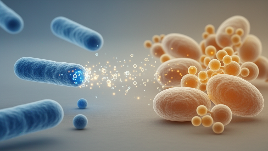
Candida albicans is a yeast most people carry without ever noticing it. It lives quietly on the tongue, in dental plaque, and on other surfaces of the mouth, normally held in check by the surrounding microbial community. But when that balance is disturbed, it can multiply aggressively, and its consequences reach well beyond simple thrush. In decayed teeth, particularly within dentin, C. albicans secretes collagenolytic enzymes that break down dentinal collagen, increasing tissue permeability and damaging the dental matrix. It also lowers the pH of its environment, promoting demineralization of hard dental tissues, and may therefore accelerate dental caries in concert with other microorganisms.
The therapeutic arsenal against this yeast is eroding. Overuse and misuse of antifungal drugs have fueled the emergence of resistant C. albicans strains, and studies have documented resistance to widely used agents such as fluconazole and itraconazole, contributing to chronic and recurrent infections. Non-albicans Candida species are increasingly recognized as additional treatment challenges. This growing resistance has pushed researchers toward alternative approaches, including postbiotic metabolites, yet studies evaluating the direct effects of postbiotic mediators on fungal biofilms have remained limited.
Probiotics, live microorganisms that can suppress the growth and colonization of C. albicans, have attracted attention for managing oral candidiasis. They produce antifungal compounds such as lactic acid and bacteriocins, and may offer benefits including reduced adverse effects compared with antifungal medications and immunomodulatory effects. But live probiotics carry drawbacks: delayed effects, variable efficacy among individuals, and an increased risk of infection in immunocompromised patients. Postbiotics sidestep these concerns. Defined as non-viable microbial preparations or their components that retain bioactive properties, postbiotic mediators are sterile, cell-free, filter-sterilized preparations containing bioactive metabolites produced by probiotic metabolic activity. Because they avoid administration of viable microorganisms, they may offer practical advantages in storage and formulation, making them particularly interesting for populations in whom live bacteria may be undesirable, although the safety and stability of each preparation require direct product-specific evaluation.
A new study published in MicrobiologyOpen investigated the antifungal effects of postbiotic mediators derived from two of the most extensively studied probiotic strains, Lactobacillus rhamnosus GG (LGG) and Lactobacillus reuteri (LR). Both strains are known for robust growth, adhesion, pathogen antagonism, and production of antimicrobial substances. The researchers assessed the mediators’ minimum inhibitory concentration (MIC) and minimum fungicidal concentration (MFC) against C. albicans ATCC 10231 following Clinical and Laboratory Standards Institute M27-A3 broth microdilution guidelines, then examined their ability to destroy mature biofilms and suppress fungal metabolic activity.
The methods were rigorous. The lactobacilli were cultured in MRS broth for 48 hours, after which cultures were centrifuged and supernatants filtered through 0.22 micrometer membranes to yield sterile cell-free preparations. The pH of each preparation was adjusted to physiological values of 7.2 to 7.4 using sodium hydroxide, a step with important consequences, since organic acids such as lactic and acetic acid exert their primary antifungal effects in their protonated, acidic form. For susceptibility testing, yeast suspensions were standardized spectrophotometrically and diluted in RPMI 1640 medium buffered with MOPS. For biofilm assays, a higher inoculum of 1.5 × 10⁶ CFU/mL was used to ensure confluent and reproducible biofilm formation within the incubation period.
The results were striking. The MIC for both LGG-derived and LR-derived postbiotic mediators was 25 percent (v/v), and no fungal growth appeared on agar at concentrations above this level, confirming the MFC value. Because the MFC equaled the MIC for both preparations, the mediators demonstrated fungicidal activity against planktonic cells. In the biofilm destruction assay, colony-forming unit enumeration showed that both mediators yielded lower viable counts than the untreated control, but the reduction was statistically significant only for the LGG group (p = 0.0038) and not for the LR group (p = 0.1537). The authors note that the absolute reduction for LGG, while significant, was relatively modest, approximately a 17.5 percent decrease relative to control, or about 0.08 log₁₀.
Scanning electron microscopy painted a vivid picture of what the treatments did to biofilm architecture. Biofilms formed on tooth slabs, fixed with glutaraldehyde, dehydrated through ascending ethanol concentrations, and coated with palladium-gold revealed reduced microbial density and fewer microcolonies on treated surfaces, with LGG-derived mediators producing the more pronounced antibiofilm effect. The XTT reduction assay, which measures metabolic activity by detecting absorbance of reduced tetrazolium compound at 490 nm, showed that both mediators significantly suppressed biofilm metabolic activity at half the MIC: mean absorbance fell from 1.78 ± 0.09 in the control to 0.96 ± 0.06 for LGG (p = 0.0009) and 1.12 ± 0.08 for LR (p = 0.0015). This indicates that the mediators exerted biologically relevant effects on biofilm physiology even though they did not achieve rapid, high-level killing of biofilm-embedded cells within the 24-hour exposure window.
The disparity between the two strains, despite comparable MIC and MFC values, may reflect strain-specific differences in metabolite composition and biofilm-targeted mechanisms. While MIC and MFC assays primarily assess fungicidal activity against planktonic cells, LGG mediators may contain metabolites such as biosurfactants, proteases, or bacteriocins that destabilize biofilm architecture by affecting extracellular matrices or hyphal formation, a critical virulence trait of C. albicans. Prior work offers clues: LGG’s major secretory peptidoglycan hydrolase Msp1 has been shown to contribute to chitin degradation in C. albicans hyphae, and lactic acid in LGG-derived preparations affected the glucan coating on hyphal surfaces, potentially making hyphae more accessible to Msp1 activity. LR, meanwhile, produces various organic acids including acetic, lactic, oxaloacetic, propanoic, decanoic, and benzoic acids, which may contribute to growth restriction or fungal cell deformation.
The study has important limitations that temper enthusiasm. Only one reference strain of C. albicans was evaluated, so the results cannot be assumed to represent the susceptibility of genetically diverse clinical isolates, antifungal-resistant strains, or non-albicans Candida species. The postbiotic mediators were tested as crude, chemically uncharacterized preparations, leaving the concentrations and relative contributions of organic acids, peptides, bacteriocin-like compounds, and other metabolites unknown; metabolomic approaches such as LC-MS/MS should be used to identify the active components. Mechanisms of action were not directly investigated, cytotoxicity and biocompatibility were not assessed in mammalian cell models, and all experiments were performed in vitro without reproducing salivary flow, host immune responses, or polymicrobial oral biofilms. The pH adjustment may also have reduced the activity of pH-sensitive organic acids, and no standard antifungal comparator such as fluconazole was included for direct benchmarking.
These caveats aside, the findings demonstrate that cell-free postbiotic mediators from LGG and LR suppress the metabolic activity of C. albicans biofilms, with LGG-derived preparations showing the greater antibiofilm effect and electron microscopy confirming altered biofilm architecture. As antifungal resistance continues to spread and dental caries remains a burden on public health, especially among children, postbiotics offer a compelling direction: the antimicrobial benefits of probiotic bacteria without the risks of administering live microorganisms. Before therapeutic or preventive applications are considered, future research must evaluate diverse clinical and resistant Candida isolates, chemically characterize the active metabolites, investigate hyphal transition and membrane integrity, assess cytotoxicity in oral epithelial and fibroblast models, and test efficacy in physiologically relevant in vivo conditions. The road from laboratory dish to dental clinic is long, but this study marks a meaningful step along it.
Subject of Research: Antifungal and antibiofilm activity of postbiotic mediators from Lactobacillus rhamnosus GG and Lactobacillus reuteri against Candida albicans
Article Title: Antifungal Activity Assessment of Postbiotic Mediators Derived From Lactobacillus rhamnosus GG and Lactobacillus reuteri Against Candida albicans
Article References: Amirinia, F., Banakar, M., Alaeddini, M., Etemad‐Moghadam, S., Pourhajibagher, M., & Noori, M. (2026). Antifungal Activity Assessment of Postbiotic Mediators Derived From Lactobacillus rhamnosus GG and Lactobacillus reuteri Against Candida albicans. MicrobiologyOpen, 15(5), Article e70439. https://doi.org/10.1002/mbo3.70439
Image Credits: AI Generated
DOI: 10.1002/mbo3.70439
Keywords: postbiotics, Lactobacillus rhamnosus GG, Lactobacillus reuteri, Candida albicans, biofilm, antifungal resistance, oral health, Antifungal, Activity, Assessment, Postbiotic, Mediators
News Source: Morgan Morrow. (October 17, 2026). Postbiotics From Probiotic Bacteria Show Antifungal Power Against Candida albicans. Scienmag.