


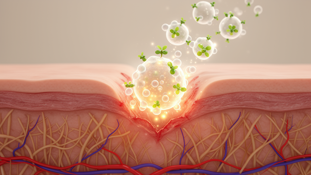
A bioactive compound isolated from the roots of Sanguisorba officinalis L., a traditional Chinese medicine long used to treat burns and scalds, has been shown to accelerate skin wound healing in mice by activating a specific immune cell pathway. The compound, ziyuglycoside I, or ZG-I, boosted the expression of a receptor called GPR34 on group 3 innate lymphoid cells within wound tissue, triggering the release of interleukin-22, a cytokine central to epithelial regeneration. The findings, published in the journal Chinese Medicine, identify GPR34 as a potential therapeutic target for skin injury-related diseases, although the work remains at the preclinical stage and has not yet been tested in humans.
The research team, led by Kewei Wang, Qingya Sun, Gang Wang, Jiayan Zhang, Yehuang Wang, Bin Jiang and Yang Zhang, set out to clarify the mechanisms by which ZG-I promotes wound healing. ZG-I is one of the bioactive compounds isolated from the roots of Sanguisorba officinalis L., an herb that has been used in China for more than 2000 years to promote hemostasis and treat burns and scalds. Previous studies had attributed wrinkle-care, antioxidant and antiviral properties to the compound, but its role in tissue repair was unknown. Group 3 innate lymphoid cells, or ILC3s, are characterized by expression of the transcription factor RORγ and are known to maintain epithelial tissue homeostasis and regeneration, yet their contribution to normal skin repair had remained largely unexplored.
To test the compound’s efficacy, the researchers created full-thickness excisional wounds, 10 millimeters in diameter, on the backs of BALB/c male mice. The animals were randomly assigned to six groups of 12: an untreated model group, a base-cream group, three ZG-I dose groups receiving 0.1, 0.2 or 0.4 milligrams per kilogram, and a positive-control group treated with recombinant human epidermal growth factor gel. Treatment was applied twice daily starting the day after surgery and continued for 14 days. The researchers then monitored wound closure rates and performed histological analyses to assess inflammation and re-epithelialization.
The results showed that topical application of ZG-I enabled faster tissue recovery compared with the base-cream group. Histopathological examination revealed markedly decreased inflammatory responses and immune cell infiltration, alongside accelerated re-epithelialization in the high-dose and positive-control groups. Masson staining demonstrated dense and well-organized collagen fiber deposition around incision sites treated with ZG-I. Immunohistochemical analysis using Ki67, α-SMA and Vimentin antibodies further showed accelerated migration, maturation and proliferation of epidermal and dermal cells in the ZG-I and positive-control groups relative to the base group, processes the authors describe as critical for effective wound healing.
Mechanistically, the team found that ZG-I upregulated the expression of GPR34 in ILC3s within wound tissues. Western blot analysis revealed a significant elevation in GPR34 levels in the skin of wounds from the high-dose group, and immunofluorescence imaging showed that the quantity of RORγt-positive, GPR34-positive cells was markedly greater than in the base group, suggesting that ZG-I promotes the activation of ILC3s. Flow cytometry analysis demonstrated that the proportion of IL-22-positive ILC3s exhibited a clear dose-dependent upward trend with increasing treatment doses, and enzyme-linked immunosorbent assays confirmed elevated secretion of IL-22 in skin tissues from the high-dose group.
Downstream signaling analysis showed that the expression ratio of phosphorylated STAT3 to total STAT3 was notably increased in the high-dose group compared with the base group, and immunofluorescence images revealed co-localization of RORγt with phosphorylated STAT3, indicating that STAT3 is significantly activated in ILC3s. Because GPR34 is a Gi/o protein-coupled receptor, the researchers also measured the expression levels and phosphorylation status of AKT, ERK, P38 and JNK in mouse skin, obtaining noteworthy results that indicate P38, AKT and ERK signaling pathways function downstream of GPR34 to regulate STAT3 activation and IL-22 expression in ILC3s. Increasing doses of ZG-I also led to a significant decrease in pro-inflammatory factors including IL-1β, IL-6 and TNF-α, and a marked increase in the anti-inflammatory factor IL-10.
In vitro experiments extended these findings. After determining the maximum non-toxic concentration of ZG-I on ILC3 cells, the researchers selected three concentrations for subsequent experiments and co-cultured the treated ILC3s with primary mouse skin fibroblasts. ZG-I significantly enhanced fibroblast viability, and both EDU-positive cell counts and migrated cell counts per field showed significant increases after drug administration. The expression of GPR34 in ILC3s rose with increasing ZG-I concentration, and flow cytometry showed significantly increased intracellular levels of IL-22 and phosphorylated STAT3, while enzyme-linked immunosorbent assays confirmed elevated IL-22 secretion. Expression levels and phosphorylation status of STAT3, AKT, ERK, P38 and JNK were also upregulated following drug administration.
To verify that GPR34 is the key receptor mediating these effects, the researchers knocked down GPR34 in ILC3s using small interfering RNA and then co-cultured the cells with mouse fibroblasts. The knockdown significantly inhibited fibroblast viability, and compared with the high-dose ZG-I group, both EDU-positive cell counts and migrated cell counts were significantly reduced in the si-GPR34 plus high-dose group, indicating that GPR34 knockdown antagonized the promoting effects of ZG-I. Levels of phosphorylated STAT3 relative to total STAT3 were also inhibited, and flow cytometry showed a decrease in the percentage of IL-22-positive and phosphorylated STAT3-positive ILC3s. The results confirmed that silencing GPR34 counteracts the impact of ZG-I in the in vitro models.
The team then tested whether ZG-I physically binds to GPR34. Molecular docking using the AlphaFold structure of mouse GPR34 predicted that the compound binds most favorably at active pocket 559, achieving the highest LibDockScore of 135.468, with key interactions including π-alkyl contacts and carbon-hydrogen bonds. A cellular thermal shift assay showed that ZG-I treatment significantly enhanced the thermal stability of GPR34, shifting the melting curve rightward, which the authors interpret as direct biophysical evidence that ZG-I engages with GPR34 within the cellular environment. Microscale thermophoresis measurements quantified the interaction, revealing that ZG-I bound wild-type GPR34 with a dissociation constant of 0.71 micromolar, while binding to a mutant GPR34 protein with key amino acid residues altered was significantly diminished, with the dissociation constant rising to 60.82 micromolar, supporting the specificity of the predicted binding pocket.
The authors acknowledge several limitations. The in vivo physiological significance of the AKT, MAPK and STAT3 signaling events has not been comprehensively established, and ILC3-specific or systemic GPR34 knockout mouse models would be needed to confirm whether ZG-I’s wound-healing effects are entirely dependent on GPR34 in vivo. The investigation of ILC3 crosstalk was confined to interactions with neutrophils and fibroblasts, leaving potential effects on macrophage polarization or T lymphocyte function unexplored, and the possibility that ZG-I acts on additional molecular targets cannot be excluded. The clinical translatability of the findings is further limited by the use of a standardized acute full-thickness wound model in healthy mice, which may not recapitulate the pathological microenvironment of chronic, non-healing wounds such as diabetic ulcers, and the study lacks long-term safety and efficacy data, including pharmacokinetic profiling and assessment of potential adverse reactions. Within these constraints, the authors conclude that ZG-I has the activity of promoting wound healing and that GPR34 can serve as a therapeutic target for ZG-I in the treatment of skin injury-related diseases.
Subject of Research: A preclinical study of ziyuglycoside I from Sanguisorba officinalis L. showing that it accelerates skin wound healing in mice by activating GPR34 signaling in group 3 innate lymphoid cells.
Article Title: Ziyuglycoside I promotes wound healing via GPR34-mediated activation of group 3 innate lymphoid cells
Article References: Wang, K., Sun, Q., Wang, G., Zhang, J., Wang, Y., Jiang, B., & Zhang, Y. (2026). Ziyuglycoside I promotes wound healing via GPR34-mediated activation of group 3 innate lymphoid cells. Chinese Medicine, 21(1), Article 212. https://doi.org/10.1186/s13020-026-01483-0
Image Credits: AI Generated
DOI: 10.1186/s13020-026-01483-0
Keywords: ziyuglycoside I, wound healing, GPR34, innate lymphoid cells, IL-22, Sanguisorba officinalis, STAT3, skin repair, molecular docking, CETSA, preclinical study, Chinese medicine
News Source: Drew Townsend. (October 30, 2026). Plant Compound Ziyuglycoside I Speeds Wound Healing by Activating Immune Cells Through GPR34 Receptor. Scienmag.



