


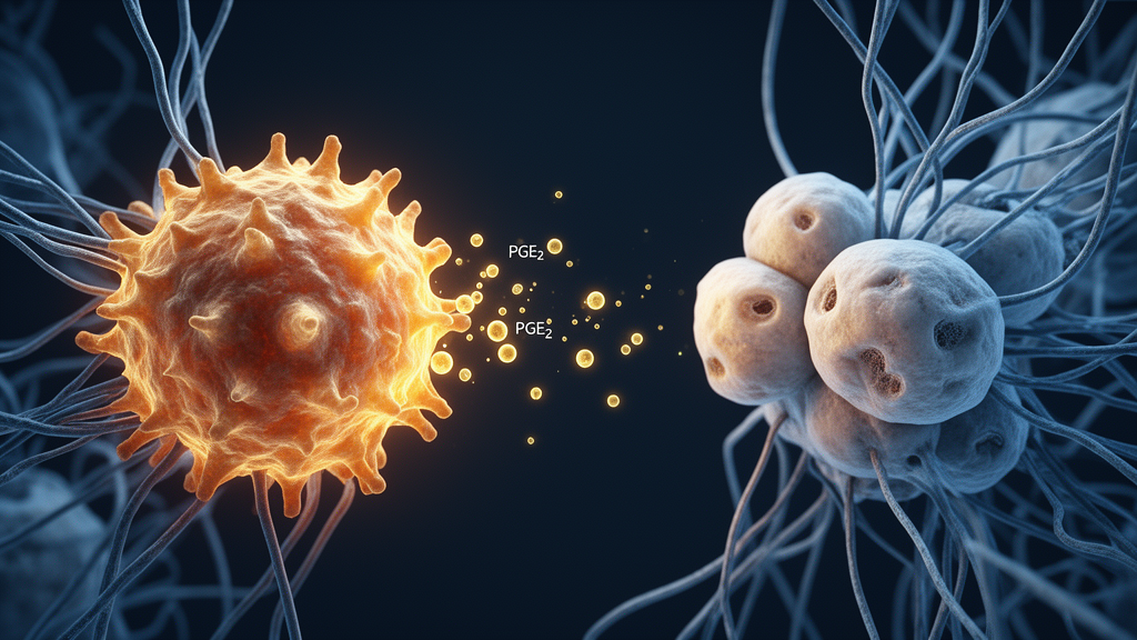
A new study published in EMBO Molecular Medicine has mapped a molecular chain of events that may explain why some cesarean section scars never heal properly, turning a routine surgical wound into a stubborn, fibrotic defect that undermines fertility. The research identifies elevated prostaglandin E2, or PGE2, as an upstream driver that pushes immune cells into a premature aging state, which then spreads to uterine fibroblasts and fuels excessive scar tissue formation. In mouse models, blocking this pathway or targeting the aging process itself significantly reduced fibrosis and restored uterine structure and function.
Cesarean section scar defect, known clinically as CSD, affects between 50 and 84 percent of women who have had a cesarean delivery, according to prior clinical studies cited in the paper. The condition involves incomplete healing of the myometrium, the muscular wall of the uterus, leaving a symptomatic niche at the incision site. Women with CSD can experience prolonged menstrual bleeding, painful periods, secondary infertility, scar pregnancy, and psychological distress. Surgical repair techniques such as laparoscopic, hysteroscopic, and transvaginal procedures can remove these niches and improve outcomes, yet some patients continue to have symptoms even after multiple operations, suggesting that unknown biological factors keep the defect active.
To uncover those factors, the team led by Yujia Yin, Huihui Chen, Weiwei Xie, Yizhi Wang, and senior authors Zhiyuan Mao, Ziyi Li, and Xipeng Wang performed quasi-targeted metabolomics on paired CSD tissues, adjacent para-CSD tissues, and healthy control myometrium. Metabolic profiling showed that both CSD and para-CSD tissues were markedly distinct from healthy controls, while showing no significant differences between each other. Prostaglandin family metabolites, key products of arachidonic acid metabolism, were significantly upregulated, with overall pathway enrichment confirmed. A follow-up targeted metabolomic analysis in an expanded cohort confirmed that PGE2, PGF2α, TXB2, and 12-HETE were significantly elevated in both CSD and para-CSD tissues compared with healthy controls, while progesterone levels showed a consistent downward trend.
The researchers then validated the enzymatic source of this PGE2 surplus. RT-PCR analysis demonstrated significant upregulation of PTGES, the gene encoding prostaglandin E synthase, the rate-limiting enzyme for PGE2 production, and PTGR1, which encodes a key enzyme in downstream PGE2 catabolism, in both CSD and para-CSD tissues. Immunohistochemistry on tissue microarrays containing 118 CSD samples and 37 controls showed robust PGES protein upregulation in both CSD and para-CSD regions. ELISA further confirmed significantly increased PGE2 levels in CSD tissues and in the serum of CSD patients. Correlational analyses revealed that PGES upregulation was significantly positively correlated with preoperative niche width and significantly negatively correlated with the thickness of residual myometrium, linking the pathway to disease severity.
To test whether elevated PGE2 actively worsens outcomes rather than merely marking them, the investigators established a murine cesarean section model in which pregnant mice at embryonic day 17 underwent bilateral longitudinal uterine incisions, with healing evaluated at eight weeks. Compared with sham-operated controls, CS-operated mice showed significantly increased fibrosis at the incision site along with elevated PGE2 levels. When the researchers gave CS mice daily intraperitoneal PGE2 injections, the animals developed significantly enhanced fibrosis, deeper and wider defects, and reduced residual myometrial thickness. Immunohistochemistry for alpha-smooth muscle actin and collagen I confirmed reduced myometrial tissue and increased fibrotic deposition. Subsequent pregnancy outcomes were also impaired, with fewer pregnancies in both uterine horns and at the CS sites, potentially resulting from reduced endometrial thickness and gland number.
Conversely, inhibiting PGE2 production produced the opposite effect. The team administered the COX-2 inhibitor celecoxib and the PGES inhibitor CAY10526 to CS mice for eight weeks. ELISA demonstrated significant reductions in PGE2 levels at the CS site in both treatment groups, accompanied by improved uterine patency rates. Histological staining confirmed reduced fibrosis at the incision site, with corresponding decreases in residual incision width and depth. The CAY-treated group showed a significant increase in residual myometrial thickness. Endometrial thickness and gland number also improved, and the number of pregnancies occurring at the CS site increased significantly at twelve weeks post-CS. Silencing PTGES using adeno-associated virus delivery of shPTGES achieved comparable benefits, including improved uterine patency, enhanced myometrial healing, reduced fibrosis, and increased endometrial thickness and gland number.
With the pathway established, the researchers turned to cellular senescence, the state in which cells stop dividing but remain metabolically active and secrete inflammatory molecules collectively known as the senescence-associated secretory phenotype. Immunofluorescence staining for the senescence marker p16 in CS mice over time revealed a peak in senescent cells at one week post-CS, before excessive fibrosis was evident, indicating that cellular senescence emerges as an early event in the fibrotic process. In PGE2-treated mice, the number of p16-positive cells remained elevated over time compared to controls. In human CSD tissues, immunohistochemistry and RT-PCR demonstrated significant upregulation of senescence and SASP markers, accompanied by reduced expression of proliferation markers and LaminB1, confirming the presence of persistent senescent cells within fibrotic niches of CSD patients.
To identify which cell types were most vulnerable, the researchers exposed human umbilical cord-derived mesenchymal stem cells, uterine smooth muscle cells, uterine macrophages, THP1-derived macrophages, and uterine fibroblasts to PGE2 in vitro. Only macrophages and uterine fibroblasts exhibited significant senescence-associated molecular features in response. Temporal analysis in the mouse model showed that PGE2-treated mice displayed a marked increase in p16-positive senescent macrophages during the early phase, one to three weeks post-CS, whereas accumulation of p16-positive senescent fibroblasts occurred predominantly during later stages. At one week post-CS, more than 40 percent of macrophages and over 30 percent of uterine fibroblasts at incision sites in PGE2-treated mice exhibited senescence, significantly higher than in controls. PGE2 treatment of THP1-derived macrophages markedly increased senescence-associated beta-galactosidase activity and significantly reduced proliferation markers Ki-67 and EdU expression, while RT-PCR revealed activation of p16 and p21-associated pathways and upregulation of SASP factors, particularly IL1B and IL8.
The critical question was how senescent macrophages communicate with fibroblasts. Conditioned medium from senescent macrophages significantly increased expression of fibroblast activation markers ACTA2, COL1A1, and TGFB, and robustly enhanced SA-beta-gal activity in uterine fibroblasts, effects that were reversed by COX-2 or PGES inhibition. Bulk RNA sequencing of PGE2-treated macrophages revealed significant enrichment of cytokine-related pathways, particularly NF-kappaB signaling. Single-cell RNA sequencing of 31,916 high-quality single cells from CSD patients and healthy uterine tissues identified three macrophage subsets, and mapping the differentially expressed genes onto these subsets revealed enrichment of IL1B-positive macrophages, which displayed robust upregulation of senescence-associated genes. Multiplex immunofluorescence confirmed a pronounced increase in senescent gamma-H2A.X-positive IL-1beta-positive macrophages in CSD tissues, spatially co-localizing with fibroblasts.
Functional validation confirmed the IL-1beta/IL-1R axis as the transmission channel. Silencing IL1B in macrophages significantly reduced IL-1beta levels in conditioned medium and attenuated both fibrotic activation and senescence induction in uterine fibroblasts during co-culture. Knockdown of IL1R in fibroblasts markedly diminished their fibrotic and senescent responses. In vivo, an anti-IL-1beta neutralizing antibody administered during the early post-injury phase significantly attenuated fibrosis, reduced CSD size, and promoted endometrial repair, while exogenous IL-1beta protein administration during the same period markedly exacerbated fibrotic remodeling. Building on this, the senolytic agent ABT263, given during the peak senescence phase of one to three weeks post-CS, markedly reduced senescent cell accumulation, decreased local PGE2 levels, and significantly improved uterine patency, fibrosis, and endometrial thickness and gland number. The senomorphic agent metformin produced similar results, reducing PGE2 expression, decreasing senescent cell abundance, improving uterine patency, and alleviating fibrosis. The authors note that these findings provide a translational rationale for targeting PGE2 signaling and senescence to improve uterine repair after cesarean section, while cautioning that metformin is pleiotropic and that premature elimination of senescent cells may compromise wound repair, underscoring the need for precisely timed intervention.
Subject of Research: The role of prostaglandin E2-driven macrophage-to-fibroblast senescence signaling in pathological fibrosis of cesarean section scar defect
Article Title: Prostaglandin E2-mediated aging microenvironment fuels pathogenic fibrosis in cesarean section scar defect
Article References: Yin, Y., Chen, H., Xie, W., Wang, Y., Qian, Y., Sheng, R., Li, D., Li, J., Zhou, X., Dai, Z., Mao, Z., Li, Z., & Wang, X. (2026). Prostaglandin E2-mediated aging microenvironment fuels pathogenic fibrosis in cesarean section scar defect. EMBO Molecular Medicine. https://doi.org/10.1038/s44321-026-00521-6
Image Credits: AI Generated
DOI: 10.1038/s44321-026-00521-6
Keywords: cesarean section scar defect, prostaglandin E2, cellular senescence, macrophages, uterine fibroblasts, IL-1beta, fibrosis, PGES, senolytics, metformin, uterine repair, mouse model
News Source: Beatrice Stafford. (October 30, 2026). PGE2 Drives a Macrophage-to-Fibroblast Aging Relay Behind Cesarean Scar Defects. Scienmag.



