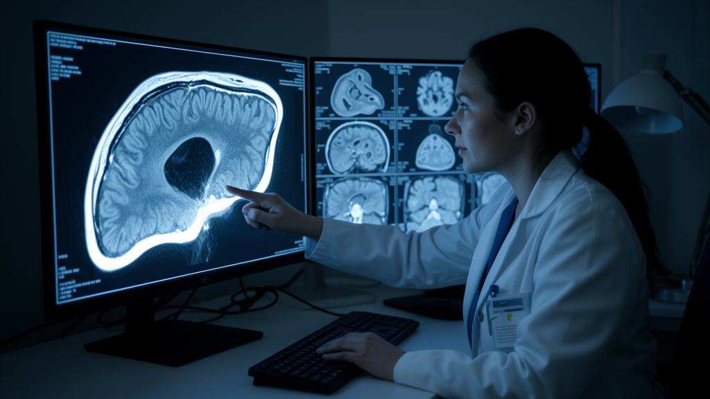
Liver surgeons face a hidden enemy they cannot see on standard scans. When a hepatocellular carcinoma is removed, pathologists sometimes find tumor cells lurking inside tiny blood vessels around the tumor, a phenomenon called microvascular invasion, or MVI. Its presence is one of the strongest predictors of poor outcomes after surgery, yet it can only be confirmed definitively by examining the resected specimen under a microscope. A new prospective study published in European Radiology Experimental reports that quantitative measurements from diffusion-weighted MRI, taken not inside the tumor but in the rim of liver tissue surrounding it, can flag this dangerous feature before the operation begins.
The research team, led by Yunfei Zhang, Jiejun Chen, Xianling Qian, and Mengsu Zeng, enrolled 95 patients with suspected hepatocellular carcinoma at Zhongshan Hospital, Fudan University, in Shanghai. After sequential exclusions for reasons such as non-HCC pathology, prior liver-directed therapy, long intervals between MRI and surgery, poor image quality, and missing pathological assessment, 70 patients formed the final cohort: 61 males, 42 of them MVI-positive, with a mean age of 55.6 years. All patients underwent MRI on a 3-T scanner using a customized multi-b-value diffusion-weighted sequence acquired during free breathing, with ten b-values ranging from 0 to 2,000 s/mm² and a total acquisition time of roughly 3.5 minutes.
What sets this study apart is where and how the measurements were made. Rather than relying on a single diffusion model, the researchers calculated voxel-wise parameters within the tumor and in automatically generated 3-millimeter and 5-millimeter peritumoral rims using five different diffusion models: the conventional monoexponential model yielding ADC, intravoxel incoherent motion (IVIM), diffusion kurtosis imaging (DKI), the stretched-exponential model (SEM), and the continuous-time random-walk (CTRW) model. Each model captures different aspects of water molecule movement in tissue, from simple cellularity to microstructural complexity and perfusion. Tumor volumes of interest were delineated by consensus by two experienced observers who were blinded to the pathological MVI status, and disagreements were resolved under the supervision of a senior abdominal radiologist with more than 30 years of experience.
The reference standard was rigorous. MVI status was determined by histopathological assessment of surgically resected specimens, defined as the presence of tumor cell nests or emboli within an endothelium-lined vascular lumen, typically located in the tumor capsule or adjacent liver parenchyma. The pathologists who made this determination were blinded to all DWI-derived parameters and model predictions, ensuring that the imaging analysis was tested against an independent ground truth.
The results revealed a striking asymmetry between the tumor interior and its surroundings. Inside the tumor, only four diffusion-coefficient-related metrics showed significant differences between MVI-positive and MVI-negative groups, all pointing toward restricted diffusion: Tumor_Mono_ADC, Tumor_DKI_Dapp, Tumor_SEM_DDC, and Tumor_CTRW_Dm, with p-values between 0.020 and 0.042. In the peritumoral regions, however, twelve parameters differed significantly between groups, including not only reduced diffusion-related indices such as P3_IVIM_Dt, P3_DKI_Dapp, P5_Mono_ADC, and P5_SEM_DDC, but also marked alterations in heterogeneity-related metrics, with elevated P3_DKI_Kapp and P5_DKI_Kapp and decreased P3_CTRW_α and P5_CTRW_α, with p-values between 0.002 and 0.042.
This pattern makes biological sense. MVI is itself a peritumoral pathological phenomenon: in MVI-positive tumors, the tumor-liver interface may harbor microscopic vascular tumor emboli, satellite nodules, microvascular remodeling, increased cellular infiltration, and reactive stromal fibrosis. These processes restrict water diffusion and enhance non-Gaussian diffusion behavior in the surrounding tissue, producing higher cell density, more intricate microarchitecture, and stronger diffusion restriction. Elevated kurtosis values and reduced CTRW alpha values both indicate enhanced tissue heterogeneity, reflecting more complex diffusion distributions at the invasive margin.
The diagnostic numbers told the same story. Among individual parameters, the best intratumoral performer was Tumor_SEM_DDC with an area under the receiver operating characteristic curve (AUROC) of 0.655. Four peritumoral parameters beat it: P3_DKI_Kapp led all individual metrics at an AUROC of 0.713, followed by P5_DKI_Kapp at 0.709, P5_CTRW_α at 0.697, and P5_IVIM_Dt at 0.664. Among clinical variables, AFP achieved an AUROC of 0.685 and tumor size 0.668. Multivariable logistic regression identified tumor size (odds ratio 1.290, 95% confidence interval 1.003–1.660, p = 0.023) and AFP level (odds ratio 3.142, 95% confidence interval 1.042–9.475, p = 0.040) as independent clinical risk factors for MVI.
After stepwise logistic regression with variance inflation factor filtering to remove multicollinear features, two independent imaging predictors survived: Tumor_SEM_DDC and P5_DKI_Kapp. Interestingly, although P3_DKI_Kapp achieved the highest individual AUROC, P5_DKI_Kapp was retained in the multivariable model, possibly because the 5-millimeter rim provided a broader and more stable representation of the peritumoral microenvironment with less redundancy relative to the other predictors. The researchers then built three models: a clinical-only model using tumor size and AFP (AUROC 0.741), an imaging-only model using the two DWI predictors (AUROC 0.759), and a combined model integrating all four variables. The combined Cli-IntraPeriDWI model achieved the best performance, with an AUROC of 0.844, sensitivity of 0.786, and specificity of 0.893. A DeLong test confirmed that this combined model significantly outperformed the clinical-only model (p = 0.039), demonstrating that the DWI features added genuine incremental value beyond tumor size and AFP alone.
To make the model’s decisions transparent rather than opaque, the team applied SHAP (SHapley Additive exPlanations) analysis. This revealed that larger tumor size, higher AFP levels, lower Tumor_SEM_DDC, and higher P5_DKI_Kapp were each associated with increased predicted MVI risk. At the individual level, the analysis showed complementary roles: P5_DKI_Kapp and Tumor_SEM_DDC contributed predominantly to MVI detection sensitivity, while AFP and tumor size played a greater role in maintaining specificity and negative predictive value. Calibration curves and a confusion matrix further supported the predictive reliability of the combined model.
The authors caution that several limitations temper these findings. The sample size of 70 patients, while prospectively powered with a minimum requirement of 62 calculated before the study began, is relatively limited, and no external validation across multiple centers or MRI systems was performed. Quantitative DWI parameters can be affected by scanner platform, field strength, gradient performance, b-value distribution, signal-to-noise ratio, respiratory motion, and model-fitting algorithms, all potential barriers to cross-platform reproducibility. The study also did not incorporate conventional qualitative imaging features such as arterial enhancement pattern, capsule integrity, and tumor margin configuration, evaluated only two peritumoral spatial scales, and did not assess inter-reader agreement because a consensus-based delineation strategy was used. Free-breathing acquisition with limited signal averaging at higher b-values may also affect parameter stability. Nevertheless, the authors suggest that incorporating peritumoral DWI analysis may enhance biomarker sensitivity not only in HCC but potentially in DWI studies of other solid tumors, offering a noninvasive route to preoperative MVI risk stratification that could inform surgical decision-making, adjuvant treatment considerations, and follow-up intensity.
Subject of Research: Preoperative prediction of microvascular invasion in hepatocellular carcinoma using peritumoral and intratumoral diffusion-weighted MRI metrics
Article Title: Preoperative prediction of HCC microvascular invasion by multimodel peritumoral diffusion MRI
Article References: Zhang, Y., Chen, J., Qian, X., & Zeng, M. (2026). Preoperative prediction of HCC microvascular invasion by multimodel peritumoral diffusion MRI. European Radiology Experimental, 10(1), Article 160. https://doi.org/10.1186/s41747-026-00817-9
Image Credits: AI Generated
DOI: 10.1186/s41747-026-00817-9
Keywords: hepatocellular carcinoma, microvascular invasion, diffusion-weighted imaging, peritumoral region, MRI, diffusion kurtosis imaging, stretched-exponential model, AUROC, SHAP, predictive model, liver cancer, radiomics
News Source: Nathaniel Bowman. (October 28, 2026). Peritumoral MRI Metrics Outperform Tumor Measures for Predicting Liver Cancer Invasion. Scienmag.