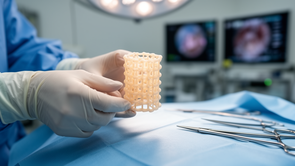
A structured review of observational studies on polyetheretherketone, or PEEK, cages used for fusion after anterior cervical corpectomy has concluded that the implants perform comparably to other fusion materials, with complication and revision rates that are not higher than those reported for competing devices. The narrative review, published in Springer Nature’s Biomedical Materials Science journal, pooled data from five retrospective studies covering 279 patients treated for degenerative cervical spinal canal stenosis. Its authors, Chris Schulz, Uwe Max Mauer and Magnus Scheer, also flag an important caveat: the pooled evidence was formally graded as low quality under the GRADE criteria, and the findings should be read with that limitation in mind.
Anterior cervical corpectomy and fusion, abbreviated ACCF, is a procedure in which one or more vertebral bodies are removed from the front of the neck to decompress the spinal canal, and the resulting gap is bridged with a fusion material. Traditionally this was an autologous bone graft, harvested from the patient, most often from the iliac crest or the fibula. Harvesting such a graft carries its own disadvantages, which is one reason alternatives have proliferated. In a survey of AOSpine members, 64 percent of surgeons said they use a cage for anterior cervical discectomy and fusion, and only 20 percent voted for autologous bone grafts; among the neurosurgeons surveyed, three-quarters preferred a cage. A survey of German and Austrian spine surgeons on ACCF specifically found that only about 16 percent still use autologous bone, while 84 percent use cages after cervical corpectomy. Thermoplastic PEEK cages have been in clinical use for ACCF for roughly 15 years, yet studies describing their clinical effectiveness and, in particular, their safety have remained rare.
To fill that gap, the authors conducted a structured literature review following the PRISMA protocol, searching PubMed/Medline, Google Scholar and the Cochrane Library for publications up to 1 July 2025 using the search terms ‘anterior cervical corpectomy + PEEK cage’. Three independent authors reviewed abstracts for eligibility. The search focused on studies that exclusively observed the clinical use of PEEK cages for fusion after cervical corpectomy in patients with degenerative cervical spinal canal stenosis, with detailed information on surgical and implant complications, material failure and revision procedures. All reported cases had to have been treated with ventral plate osteosynthesis in addition to cage fusion, and at least one clinical and one radiological outcome parameter had to be presented. Studies on biomechanical and animal model testing were excluded, as were studies with fewer than five cases, clinical courses shorter than six months, reviews, editorials, commentaries and conference abstracts or posters without accessible demographic and clinical outcome data. Where duplicate publications from the same group existed, the study with the most cases and the most detailed data was selected.
Only five publications, together documenting 279 cases, met the inclusion criteria. All five were nonrandomised, non-controlled, retrospective observational studies published between 2014 and 2019 in Europe and the USA. To objectify the risk of bias, the authors applied a modified version of the National Heart, Lung, and Blood Institute Quality Assessment Tool for case series studies, supplementing the original rating with questions on prospective study design and drop-out reporting. Under this modified eleven-criterion assessment, four of the five studies were rated of good quality and one of fair quality. Statistical analyses were performed with Open Meta [Analyst], with heterogeneity assessed using the I² statistic and considered substantial when it exceeded 50 percent at a P-value below 0.05. Weighted averages and 95 percent confidence intervals were calculated for stable fusion rates, severe subsidence, surgical access complications, implant complications and revision surgeries, and the evidence was classified according to the GRADE criteria.
The 279 patients included 102 women, or 37 percent, and 177 men, or 63 percent, with a median patient age between 51 and 65 years across the studies. Corpectomies of one vertebral body were performed in 199 patients, or 71.3 percent; corpectomies of two vertebral bodies in 68 patients, or 24.4 percent; and corpectomies of three vertebral bodies in 12 patients, or 4.3 percent. Follow-up intervals across the pooled publications ranged from six months to six years. The authors note that the clinical and radiological outcome parameters of the included studies were heterogeneous and the follow-up intervals inconsistent, which constrained the analysis.
On clinical outcomes, neck pain was reported to have improved postoperatively in four of the five studies, with no data available in one. A reduction in visual analogue scale pain scores of 49 percent was determined from two of the five studies, ranging from 43.5 to 53.5 percent. A 51 percent reduction in Neck Disability Index scores was demonstrated, calculated from two of five studies, with reductions ranging from 40 to 62 percent. The severity of myelopathy improved postoperatively in all five studies, albeit with inconsistent parameters, and a reduction in the European Myelopathy Score of 17.5 percent was observed, determined from three of five studies and ranging from 12 to 23 percent. Radiologically, the stable fusion rate was reported in all included studies, ranging between 70 and 100 percent. Stable fusion was observed in 215 of 244 patients during the observation period, and the weighted average stable fusion rate was 0.88, with a 95 percent confidence interval of 0.75 to 1.02 and moderate heterogeneity between studies of I² = 48.75 percent. Only three of the five studies provided information on postoperative changes in the sagittal profile of the cervical spine; in those, the sagittal profile improved in the majority of patients, the regional kyphosis rate decreased significantly, and the mean increase in the C2–C7 lordosis angle ranged from 1.5 to 5.1 degrees depending on the number of vertebral bodies replaced.
Complications typical of this surgical access were reported in all included studies and observed in 20 of 279 patients, with rates ranging between 3.96 and 11.43 percent. The weighted average surgical access complication rate was 0.07, with a 95 percent confidence interval of 0.04 to 0.11 and moderate heterogeneity of I² = 45.31 percent. In nine of the 20 patients with complications, surgical revision was necessary; the most common diagnosis was hematoma, accounting for six of the 20 cases, all of which were surgically revised. Postoperative implant failure exclusively involved loosening or breakage of the osteosynthesis screws and was observed in 13 of 247 patients, ranging between 0 and 15.71 percent across studies. The weighted average implant failure rate was 0.05, with a 95 percent confidence interval of −0.02 to 0.12 and substantial heterogeneity of I² = 98.31 percent. In five of the 13 affected patients, or 38.5 percent, revision was performed because of the screw loosening or breakage. Breakage of the ventral plates or of the PEEK cages themselves was not detected in any case. Information on cage subsidence was published in only four of the five studies; the subsidence rate ranged from 12 to 76.2 percent and the subsidence distance from 1 to 6 millimetres. Severe subsidence, defined as more than 3 millimetres, was found in 25 of 209 cases, ranging between 8.16 and 20 percent, with a weighted average severe subsidence rate of 0.11, a 95 percent confidence interval of 0.06 to 0.18 and substantial heterogeneity of I² = 64.42 percent.
Revision surgery for a fusion complication, with or without dislocation of the ACCF construct, was necessary in 10 of 247 cases, with rates across studies ranging between 0 and 10 percent. The weighted average revision rate was 0.04, with a 95 percent confidence interval of 0.00 to 0.08 and substantial heterogeneity of I² = 88.69 percent. In eight of the 10 cases, the fusion problem occurred after multilevel ACCF, and all dislocations requiring revision occurred within the first four months after surgery, with eight of the 10 patients affected within the first eight weeks. The most common reason for revision was a fracture of a screw-bearing vertebral body with subsequent ventral dislocation of the cage-screw-plate complex: the caudal vertebral body was involved in five of the 10 cases, the cranial fusion vertebra in one, and both vertebral bodies in two. In four of the five patients with such a fracture, osteoporosis was also present, and in three of the five the revision was immediately preceded by an acute fall. Three of the 10 patients showed dorsal dislocation, all of them smokers with diabetes mellitus and osteoporosis; notably, these cases came from a single publication in which the implant system apparently lacked a connector screw between the cage and the ventral plate, a feature present in the systems used in the other four studies, where no dorsal dislocation occurred. The authors suggest this connecting screw could prevent dorsal migration of the cage. In two further patients, revision was performed on a rather prophylactic indication, upon detection of cage subsidence combined with screw loosening even without translational dislocation.
Placed against the wider literature, the review’s numbers sit within the range reported for other fusion materials. Biomechanical tests have shown no significant differences in primary stability between iliac crest grafts, titanium and PEEK cages, and a review summarised by the authors found no relevant differences in neurological outcomes between bone and titanium cage fusion in the first two years after ACCF. In that literature, dislocations were found in 4.9 percent of cases for bone and titanium implants, the pseudarthrosis rate was 8.5 percent, and implant failure was observed in 5.4 percent of cases. Fusion complications tend to occur more frequently with bone autografts and allografts, at 25 to 40 percent, than with cages, although a distractable titanium cage has been associated with fusion rates around 50 percent and severe subsidence of 31.4 percent in some studies. A meta-analysis of 2005 cases across 34 papers focusing on cage subsidence after ACCF found an average severe subsidence rate of 7 percent across all materials, with the highest rates for allograft bone at 27 percent and static titanium cages at 10 percent, followed by PEEK cages at 5 percent. The review’s weighted average stable fusion rate of 88 percent falls within the published range for titanium cages or bone ACCF, and its 4 percent revision rate is not increased relative to competing materials, and even tends to be lower than reported postoperative material failure of 6 percent for single-level and up to 75 percent for multilevel ACCF with bone and titanium cages.
The authors are careful about the strength of these conclusions. According to the GRADE quality criteria, the evidence from the pooled data was formally classified as low, downgraded because of the inconsistent use of radiological and neurological examination parameters across studies and discrepancies in follow-up intervals. No raw data were made available in the included studies, the information on results is partially incomplete and highly heterogeneous, and the caseload in some individual studies is very low. One included study also does not consider a completely uniform patient population, incorporating a few cases with tumours, trauma and inflammation. Individual studies reached contradictory conclusions: two reporting a 100 percent fusion rate recommended the PEEK cage system, while another reporting a fusion rate of only 70 percent, with fusion complications requiring revision in 2 of 20 cases, advised against it. Comparative studies of fusion materials remain rare and their results mixed, and reasons for rejecting PEEK implants cannot be reliably deduced from the current literature. The authors identify older osteoporosis patients after multilevel ACCF with an increased tendency to fall as a risk group for ventral implant dislocation, and suggest that fall risk assessment, cervical orthoses for the first four months after surgery, fall prevention, anti-osteoporotic therapy and, where clinically appropriate, preoperative improvement of bone quality may be useful measures, while noting that bracing, fall prevention and bone health optimisation are not yet standard practice for ACCF. They conclude that further insights with strong evidence can only be obtained through randomised controlled trials with prospective comparisons of the various materials using homogeneous clinical, neurological and radiological outcome parameters.
Subject of Research: Safety and effectiveness of PEEK cages for fusion after anterior cervical corpectomy in degenerative spinal stenosis
Article Title: Safety and effectiveness of PEEK cages for fusion after anterior cervical corpectomy in patients with degenerative spinal stenosis: a narrative review
Article References: Schulz, C., Mauer, U. M., & Scheer, M. (2026). Safety and effectiveness of PEEK cages for fusion after anterior cervical corpectomy in patients with degenerative spinal stenosis: a narrative review. Biomedical Materials Science, 1(1), Article 3. https://doi.org/10.1186/s44495-025-00002-x
Image Credits: AI Generated
DOI: 10.1186/s44495-025-00002-x
Keywords: PEEK cages, anterior cervical corpectomy and fusion, degenerative spinal stenosis, spinal fusion, cage subsidence, implant dislocation, osteoporosis, revision surgery, systematic review, GRADE evidence, spine surgery, fusion materials
News Source: Ophelia Keating. (October 29, 2026). PEEK Cages Show Low Revision Rates After Cervical Corpectomy, Review Finds. Scienmag.