Canine gastric carcinoma, one of the rarest yet most lethal malignancies in dogs, has long frustrated veterinary oncologists with its insidious onset and devastating prognosis. A new study published in the journal Veterinary Oncology has now delivered the most detailed picture yet of how these tumors behave at the microscopic level, and why so many affected dogs survive only weeks after diagnosis. By examining full-thickness gastric biopsy specimens from 40 dogs, a research team led by veterinary pathologists at the Royal Veterinary College in the United Kingdom has mapped the architectural and immunological features of the disease with a precision that superficial endoscopic biopsies simply cannot match. The findings carry implications not only for companion animal medicine but also for the broader understanding of gastric cancer biology shared between dogs and humans.
The scale of the diagnostic challenge is difficult to overstate. Canine gastric carcinoma accounts for roughly eight percent of gastrointestinal tumors in dogs and less than one percent of all canine neoplasms, yet it is typically diagnosed in older animals with a median age around eight to nine and a half years. Breeds frequently affected include Golden Retrievers, Boxers, Staffordshire Bull Terriers, Labrador Retrievers, Border Collies, and Belgian Shepherds, with Belgian Shepherds and Norwegian Lundehunds the only breeds suspected of a genuine genetic predisposition. Clinical signs are subtle and often mistaken for ordinary gastrointestinal upset, which delays presentation. By the time a diagnosis is made, metastatic disease is common, and survival times frequently fall below three months. Even when surgery is performed, median survival hovers around six months, with adjuvant chemotherapy offering some improvement. Because imaging findings are non-specific and endoscopic biopsies sample only the mucosal surface, many cases evade definitive diagnosis until the disease is far advanced.
The new study addressed a critical gap: the biological meaning of microscopic tumor architecture and the immune microenvironment in this cancer. Using 40 full-thickness gastric biopsies collected by surgical excision between 2009 and 2019, the team evaluated a battery of histomorphological parameters adapted from human gastric cancer pathology. These included mitotic count, depth of tumor infiltration, intravascular invasion, tumor budding, total stromal percentage, and tumor-infiltrating lymphocytes. They also performed immunohistochemistry for programmed death-ligand 1, the immune checkpoint molecule that in human gastric cancer serves as both a prognostic biomarker and a predictor of response to immune checkpoint blockade therapy. A deep-learning, single-cell analysis pipeline applied to whole-slide scanned images provided an additional, algorithm-based measure of lymphocyte distribution, validated against pathologist annotations.
The results paint a portrait of a remarkably invasive malignancy. Only one tumor in the cohort was confined to the submucosa; eleven invaded into the muscularis layer, sixteen reached the serosa, and twelve extended into the omentum. Intravascular invasion, the presence of tumor cell emboli within blood or lymphatic vessels, was detected in 55 percent of cases and was positively correlated with depth of infiltration, suggesting that as tumors burrow deeper into the gastric wall they increasingly seed the vasculature. Tumor budding, defined as isolated single carcinoma cells or clusters of up to four cells at the invasive front, was frequent, with a median of 18 buds per hotspot field and more than two-thirds of cases classified as high budding. These features mirror the aggressive behavior documented in human gastric adenocarcinoma, where budding and vascular invasion are recognized hallmarks of metastatic potential.
Histological classification using the World Health Organization and Lauren schemes revealed that undifferentiated carcinoma predominated, accounting for half of all cases, followed by signet ring, tubular, and mucinous subtypes, with a single papillary case. Interestingly, only the WHO classification significantly stratified survival in Kaplan-Meier analysis, with tubular carcinoma showing the shortest median survival of just one day, while the Lauren system failed to discriminate outcomes. This contrasts with human gastric cancer, where diffuse and signet ring subtypes typically carry the worst prognosis, and with earlier canine reports that identified diffuse morphology as most common. The authors caution that uneven subtype representation and small cohort size likely temper these prognostic comparisons, underscoring how much remains unknown about the clinical relevance of histological subtyping in dogs.
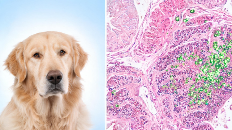
The immunohistochemical findings were striking. PD-L1 expression was detected in all 40 tumors, with labeling predominantly cytoplasmic rather than membranous, a pattern increasingly recognized across canine epithelial, round cell, and melanocytic neoplasms. When compared within the same host, neoplastic gastric epithelium showed significantly higher PD-L1 expression than adjacent normal mucosa, where labeling was infrequent and weak. The median percentage of immunolabeled tumor cells was 50 percent, and combining the percentage of positive cells with staining intensity produced a median PD-L1 score of 80. Notably, PD-L1 score correlated moderately with mitotic count, hinting at a possible link between cellular proliferation and immune evasion, as has been proposed in human gastric cancer where PD-L1 upregulation suppresses antitumor T-cell responses.
The tumor microenvironment added another layer of complexity. Tumor-infiltrating lymphocytes were identified at the invasive front, within the intratumoral stroma, and in peritumoral gastric tissue, with a median combined value of 19 percent. Total stromal percentage, a measure of the fibrous tissue accompanying the tumor, showed a strong positive correlation with lymphocyte density, suggesting intimate crosstalk between stromal architecture and immune cell recruitment. The authors speculate that cancer-associated fibroblasts, through transforming growth factor beta-mediated deposition of dense extracellular matrix, may shape this inflammatory landscape, potentially creating physical barriers that restrict effective T-cell infiltration even as lymphocytes accumulate. Intriguingly, PD-L1 expression did not correlate with lymphocyte abundance, implying that checkpoint activation may restrain antitumor immunity independently of how many immune cells are physically present.
When survival was analyzed, only one parameter emerged as consistently associated with outcome: the PD-L1 score. In univariable Cox proportional hazards regression, PD-L1 score was significantly associated with shorter survival, with a hazard ratio of 1.009 per unit score and an area under the receiver operating characteristic curve of 0.746. Using a cut-off of 60, dogs with high PD-L1 expression survived a median of 27.5 days compared with 77 days for those with low expression. Tumor budding showed a moderate negative correlation with survival duration but did not retain significance in regression analysis, while mitotic count, depth of invasion, intravascular invasion, stromal percentage, and lymphocyte measures all failed to predict outcome independently. The median survival across the whole cohort was a sobering 20 days, reflecting the reality that many dogs are euthanized at or shortly after diagnosis on welfare grounds, a factor the authors acknowledge may confound associations with tumor biology.
The study is not without limitations, which the authors confront candidly. The modest sample size relative to the number of variables analyzed raises the risk of statistical overfitting, making the survival analyses explicitly exploratory. Survival data were unavailable for nine of the 40 dogs, subtype representation was uneven, and the absence of a large independent normal cohort limits interpretation of whether cytoplasmic PD-L1 represents pathological upregulation or physiological baseline. Nonetheless, the use of full-thickness sections represents a substantial methodological strength, enabling comprehensive assessment of invasion depth and tumor-stromal interactions that superficial endoscopic biopsies cannot provide. This design choice reduced the sampling bias that has hampered previous histological studies of the disease.
The broader significance of the work lies in its convergence with comparative oncology. Dogs spontaneously develop gastric carcinomas that share molecular and histological features with the human disease, including disruptions of E-cadherin and beta-catenin signaling, epithelial-mesenchymal transition markers such as vimentin, and aberrant expression of cell adhesion molecules like EpCAM. The demonstration that PD-L1 is widely expressed in canine gastric carcinoma and associates with poor survival opens a plausible avenue for immune checkpoint-based approaches in veterinary medicine, where anti-PD-L1 antibodies have already shown clinical benefit in dogs with metastatic melanoma. Validation in larger, prospectively collected cohorts with complete clinical follow-up will be essential, but the message of this study is clear: in one of the deadliest cancers of man’s best friend, the immune escape machinery of the tumor, not its architecture alone, may hold the key to prognosis and, eventually, to treatment.
Methodologically, the study benefited from standardized approaches borrowed from human gastrointestinal pathology. Mitotic counts were enumerated across ten high-power fields measuring 2.37 mm², a convention that allows comparison with published canine reference intervals, while invasion depth was graded on a four-tier scale extending from submucosa to omentum. Immunolabeling was performed on formalin-fixed, paraffin-embedded sections using an autostainer platform after heat-induced antigen retrieval, techniques that support reproducibility across diagnostic laboratories.
The diagnosis date served as the reference point for survival calculations because it was the only time point reliably documented across all contributing practices, a pragmatic choice that reflects the realities of retrospective multicenter veterinary studies. Questionnaires distributed to referring practices supplemented medical records from the two academic hospital cases, illustrating how collaboration between commercial diagnostic laboratories and veterinary schools can assemble cohorts for rare cancers that no single institution could accumulate alone.
The cytoplasmic staining pattern observed here also carries practical significance for veterinary diagnostics. Unlike human clinical algorithms, which rely on membranous PD-L1 scoring refined through clinical trials, no validated scoring system yet exists for canine tumors, and cytoplasmic labeling complicates direct translation of human thresholds. As anti-PD-1 and anti-PD-L1 agents enter veterinary use, correlating staining patterns with treatment response in naturally occurring cancers such as gastric carcinoma may help define which patients are most likely to benefit, while simultaneously informing whether dogs can serve as relevant preclinical models for human gastric cancer immunotherapy research.
Subject of Research: Histomorphological features and PD-L1 expression in canine gastric carcinoma assessed using full-thickness biopsy specimens
Article Title: Histomorphological features and PD-L1 expression in canine gastric carcinoma using full thickness biopsy
Article References: Lean, F. Z. X., Tibbles, M. L., Hulme, B., Erles, K., Pittaway, R., Pittaway, C., Priestnall, S. L., Castillo, S. P., & Hardas, A. (2026). Histomorphological features and PD-L1 expression in canine gastric carcinoma using full thickness biopsy. Veterinary Oncology, 3(1), Article 20. https://doi.org/10.1186/s44356-026-00074-3
Image Credits: AI Generated
DOI: 10.1186/s44356-026-00074-3
Keywords: canine gastric carcinoma, PD-L1, tumor budding, tumor-infiltrating lymphocytes, tumor stromal percentage, immunohistochemistry, veterinary oncology, mitotic count, intravascular invasion, WHO classification, immune checkpoint, dog cancer
Cite Scienmag News
APA
MLA
Chicago
Nathaniel Bowman. (September 3, 2026). PD-L1 Emerges as Key Survival Marker in Aggressive Canine Gastric Cancer. Scienmag. https://scienmag.com/pd-l1-emerges-as-key-survival-marker-in-aggressive-canine-gastric-cancer/
Nathaniel Bowman. “PD-L1 Emerges as Key Survival Marker in Aggressive Canine Gastric Cancer.” Scienmag, 3 September 2026, https://scienmag.com/pd-l1-emerges-as-key-survival-marker-in-aggressive-canine-gastric-cancer/. Accessed 3 September 2026.
Nathaniel Bowman. “PD-L1 Emerges as Key Survival Marker in Aggressive Canine Gastric Cancer.” Scienmag. September 3, 2026. https://scienmag.com/pd-l1-emerges-as-key-survival-marker-in-aggressive-canine-gastric-cancer/
Copy citation
Download RIS
Tags: breed susceptibility to canine gastric tumorscanine gastric carcinomacanine gastric tumor diagnosiscomparative gastric cancer biologydog cancergastric biopsy in dogsimmune checkpointimmunohistochemistryimmunological features of canine gastric cancerintravascular invasionmicroscopic tumor architecture in dogsmitotic countPD-L1PD-L1 as survival marker in dogsprognosis of canine gastric carcinomatreatment implications for canine gastric cancertumor buddingtumor stromal percentagetumor-infiltrating lymphocytesveterinary cancer researchveterinary oncologyveterinary oncology gastric cancerWHO classification