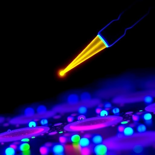
In the race to unlock the intricacies of individual cells, a technology once confined to the realm of physics laboratories is now quietly transforming biomedicine. Optoelectronic tweezers, or OETs, have evolved from a niche optical manipulation trick into a versatile, programmable platform capable of catching, moving, sorting, and interrogating single cells with unprecedented gentleness and scale. Unlike traditional tools that rely on physical probes, labeled targets, or microfluidic traps that can shear delicate membranes, OETs exploit beams of light projected onto a semiconductor chip to sculpt electric fields that push, pull, and corral cells without ever touching them. The secret is a phenomenon called optically induced dielectrophoresis, in which light patterns cast onto a photoconductive material become virtual electrodes, generating precise forces that can manipulate everything from a single bacterium to a tumor cell, all while leaving the cells viable and ready for downstream analysis.
At the heart of an OET system lies an elegantly simple sandwich: a liquid layer containing the cells is placed between a transparent conductive top plate and a bottom substrate coated with a photoconductive material, typically amorphous silicon. In darkness, the photoconductor behaves as an insulator, and the electric field across the liquid chamber remains uniform and weak. Shine a focused pattern of light onto that layer, however, and the illuminated spots suddenly become highly conductive, acting exactly like addressable microelectrodes. The resulting nonuniform field exerts a dielectrophoretic force on any polarizable particle or cell in the vicinity, allowing a computer-controlled projector to instantly reconfigure the trapping zones into any desired geometry. This virtual electrode concept is the game‑changer: there are no physical electrodes to fabricate, clog, or clean, and the manipulation patterns can be updated at video rates, meaning thousands of cells can be handled in parallel while continuously monitoring their responses under a microscope. Additional electrokinetic effects such as AC electroosmosis and electrothermal flow can also be harnessed or suppressed by tuning the frequency and amplitude of the AC voltage, giving researchers an astonishing degree of command over the cellular microenvironment.
What turns this physical principle into a biomedical revolution is the sheer adaptability of the platform. Early OET devices used a conventional digital micromirror device projector and an amorphous‑silicon chip, but the past five years have witnessed an explosion of innovations. Grayscale imaging, LCD‑based illumination, and laser‑interference patterns now allow pixel‑level control of light intensity, smoothing out electric field gradients to avoid stressing sensitive cells. The photoconductive layer itself has undergone a makeover: coating it with polyethylene glycol brushes or supported lipid bilayers virtually eliminates nonspecific cell adhesion, while dual‑layer and phototransistor‑based designs push the resolution below the diffraction limit, making it possible to trap and rotate individual virus particles or stretch single DNA molecules. New materials such as organic photoconductors dramatically lower fabrication costs and even enable flexible, disposable chips. Engineers have also broken free from the classic vertical‑field geometry, devising lateral‑field OETs and hybrid vertical–lateral schemes that generate dielectrophoretic forces in more directions, enabling cell rotation, rolling, and three‑dimensional positioning. Self‑powered OETs that harvest energy from ambient light or integrated photovoltaic cells further reduce the hardware footprint, opening the door to portable, point‑of‑care devices that could one day operate in a doctor’s office rather than a dedicated cleanroom.
The real power of OET becomes apparent when it stops merely pushing cells around and begins to merge with other single‑cell technologies. By integrating light‑controlled electroporation capabilities into the same chip, researchers can first nudge a target cell to a chosen location and then apply a precisely timed voltage pulse to open transient pores in its membrane, delivering DNA plasmids, mRNA, or CRISPR components into the cytoplasm with efficiencies that rival viral vectors while avoiding the safety concerns of viral transduction. Coupled with on‑chip cell lysis, the platform can then release intracellular contents—proteins, metabolites, and RNA—immediately into a microfluidic channel for capture and analysis. This tight choreography eliminates the sample loss and contamination that plague workflows where each step happens in a separate tube or machine, making OET an ideal hub for preparing single cells for next‑generation sequencing or mass spectrometry. Fluorescence and Raman spectroscopy modules can be overlaid directly on the manipulation zone, so that as a cell is being held in a virtual trap, its spectral fingerprint is recorded label‑free, revealing metabolic states, membrane lipid composition, or drug accumulation kinetics without any chemical label.
Microfluidics and OET form a particularly symbiotic pair. Flowing cells into an OET chip through a network of microchannels enables high‑throughput screening and sorting akin to fluorescence‑activated cell sorting, but with the critical advantage that cells can be inspected and re‑inspected under multiple conditions before a decision is made. A rare circulating tumor cell, for example, can be shuttled back and forth between a culture chamber and an imaging window, exposed sequentially to different chemotherapeutic agents, and its viability monitored in real time before being directed into a collection well for single‑cell sequencing. When the manipulation patterns are combined with electrowetting‑on‑dielectric or optoelectrowetting, the system acquires the ability to manipulate not just cells but also droplets, meaning that a selected cell can be encapsulated inside a picoliter‑volume aqueous compartment and then merged, split, or incubated with reagents, essentially creating a programmable digital microfluidic lab. This droplet‑based extension is what allows a single B lymphocyte to be isolated, its antibody secretion trapped inside the droplet, and the binding activity of that antibody measured against a panel of antigens—all within minutes and without the cell ever leaving the field of view.
Artificial intelligence and deep‑learning algorithms have recently been grafted onto OET control systems, transforming them from manually operated instruments into autonomous hunters of rare cells. Neural networks trained on bright‑field and fluorescence images can recognize cell types, distinguish live from apoptotic cells, or detect subtle morphological changes associated with drug response up to ten times faster than a human operator. Once a target is identified, reinforcement‑learning algorithms compute collision‑free paths for hundreds of cells simultaneously, orchestrating a high‑speed ballet in which cells are sorted into different reservoirs at rates exceeding thousands per hour. Closed‑loop tracking ensures that if a cell drifts out of its assigned trap due to fluid flow or Brownian motion, the light pattern instantly adapts to recapture it. This marriage of machine learning and optofluidics not only boosts throughput but also eliminates operator bias, making large‑scale single‑cell studies statistically robust and reproducible.
While impressive in the lab, OET’s most compelling promise lies in real‑world translation, and nowhere is this more evident than in the Beacon platform, a commercial photoconductive system that is already reshaping the pipelines of pharmaceutical companies. In antibody discovery, B cells harvested from immunized animals or human donors are loaded onto the chip, and each cell is individually encapsulated in a nanowell or droplet. The secreted antibodies are then screened against fluorescently labeled target antigens, and the system rapidly identifies those rare cells producing high‑affinity, blocking antibodies. Crucially, the same OET force that holds the cell during screening can then gently lift it out of the array and export it into a microtiter plate for single‑cell reverse transcription and PCR, enabling the heavy‑ and light‑chain genes to be sequenced and cloned. This workflow compresses a process that once took months down to a few days, providing a direct link from functional screening to recombinant antibody production and accelerating the development of therapeutic candidates against emerging infectious diseases and cancers.
Cell line development, a critical bottleneck in biomanufacturing, has similarly benefited from OET‑based automation. Establishing a stable, high‑yielding Chinese hamster ovary (CHO) cell clone for producing a recombinant protein often requires screening thousands of single‑cell clones over many weeks. On an OET chip, individual cells can be deposited into growth‑promoting microwells, and their bright‑field images can be captured every few minutes to document the precise moment of cell division, thereby proving clonality beyond any doubt—a regulatory requirement. The same integrated imaging system can simultaneously quantify fluorescent reporter proteins or intracellular metabolite indicators, ranking clones by productivity and growth rate without disturbing the culture. Once top candidates are identified, OET patterns can be used to retrieve the chosen colonies, bypassing the laborious and contamination‑prone steps of manual colony picking. The result is a dramatically shortened timeline from transfection to master cell bank, with an auditable digital record of every single cell’s pedigree.
In the burgeoning field of adoptive cell therapy, where a patient’s own T cells are engineered to fight cancer, OET provides a window into the functional heterogeneity that determines clinical success. Using light‑patterned traps, researchers can create precisely controlled microenvironments in which a single cytotoxic T lymphocyte is brought into contact with a tumor cell, and the ensuing lytic attack can be observed in real time. Fluorescent reporters for granzyme B, perforin, or caspase activation inside the target cell yield a direct readout of killing efficiency, while secreted cytokines such as interferon‑gamma and IL‑2 can be captured by bead‑based sensors positioned just micrometers away. Because the system can screen thousands of such one‑on‑one encounters in parallel, it becomes feasible to identify those rare T‑cell clones with superior polyfunctionality or to validate the specificity of T‑cell receptors against patient‑derived neoantigen‑presenting cells, guiding the selection of the most potent effectors for infusion.
Beyond immune cell killing, OET’s label‑free physical measurement capabilities open a fresh dimension of single‑cell characterization. By maneuvering a single cell relative to a fixed reference point and carefully analyzing its motion in response to a known dielectrophoretic force, researchers can extract its mass, density, membrane capacitance, and cytoplasmic conductivity. These parameters act as intrinsic biophysical markers that reflect the cell’s state: a cancer cell undergoing apoptosis exhibits a characteristic drop in membrane capacitance, while a stem cell differentiating toward a myocyte shows a measurable shift in density. Unlike biochemical assays, these measurements require no antibodies, no dyes, and no genetic modification, making them especially attractive for cells that will be reinfused into a patient. Moreover, OET can stretch and twist single biomolecules such as DNA and polysaccharides, providing insights into protein‑DNA interactions, replication stalling, and drug‑induced crosslinking that complement classical biochemistry.
Despite all these advances, barriers remain before OET can become a ubiquitous laboratory appliance. The capital cost of a commercial system, with its precision optics, high‑voltage amplifiers, and cleanroom‑fabricated photoconductive chips, still puts it out of reach for many academic labs. The most common photoconductor, amorphous silicon, requires deposition conditions and lithographic steps that cannot be easily replicated outside a dedicated nanofabrication facility; thus, each chip becomes a consumable that must be sourced from a handful of suppliers. Although solution‑processable organic semiconductors promise cheap, printable alternatives, their photoconductivity and surface roughness yet lag behind silicon, leading to lower cell viability and weaker manipulation forces. A related challenge is standardization: a voltage and frequency combination that works beautifully for a Jurkat T‑cell line may be completely inappropriate for primary hepatocytes or neural stem cells, yet no comprehensive database of OET parameters for different cell types exists, forcing every new user to spend weeks optimizing conditions from scratch.
Another friction point is the gap between the pristine buffers used in device development and the messy reality of clinical samples. Whole blood, bone marrow aspirates, or tumor dissociates contain debris, red blood cells, and platelet aggregates that can foul electrodes and scatter light unpredictably. Robust OET operation therefore demands integrated, automated sample preparation modules capable of lysing red blood cells, filtering aggregates, and pre‑enriching target cells before they enter the manipulation zone. The field is moving toward fully enclosed, cartridge‑based systems that accept a raw patient specimen and output sorted, sequenced, or genetically edited cells, but such systems require multidisciplinary engineering that few groups have yet mastered. In vivo applications, where OET might one day manipulate circulating cells within blood vessels or guide therapeutic stem cells to sites of injury, remain squarely in the realm of speculation because the photoconductive chip and external light source are incompatible with deep‑tissue regions and immune responses to implanted materials.
Looking ahead, the trajectory of OET is one of convergence and democratization. Researchers are exploring perovskite‑based photoconductors that combine high photoconductivity, low processing temperature, and tunable spectral response, which could lead to cells being manipulated with specific wavelengths that minimize phototoxicity. The integration of OET with high‑speed confocal and light‑sheet microscopy will allow three‑dimensional tracking and manipulation of cells within thick hydrogels, mimicking the architecture of real tissues. Simultaneously, the fusion of OET with single‑cell multi‑omics—where a single cell’s transcriptome, proteome, and metabolome are captured on the same chip—will provide a breathtakingly rich description of the molecular states that underlie the biophysical signatures already measurable. Efforts to standardize parameters through open‑source databases and shared control algorithms, coupled with the introduction of portable, low‑cost OET kits that attach to ordinary smartphone cameras, may soon place this technology in the hands of field epidemiologists and rural clinicians, enabling on‑the‑spot characterization of immune responses during an outbreak.
In time, the dictum that biology must be studied at the population level will no longer hold; OET is a cornerstone of the emerging single‑cell era, a conduit through which light is transformed into a scalpel, a conveyor belt, and a microscope all at once. As photoconductive materials become cheaper, artificial intelligence becomes smarter, and the integration with microfluidics and omics becomes seamless, these optical tweezers are poised to escape the confines of specialist laboratories and become as routine as a flow cytometer or a PCR machine. The journey from a laser‑drawn virtual electrode to a single‑cell therapy that saves a life is no longer a science‑fiction arc but an engineering roadmap being assembled one photon at a time, promising to reshape how we discover antibodies, manufacture biologics, and engineer the immune system against cancer.
Subject of Research: Optoelectronic tweezers for single-cell manipulation, functional analysis, and translational applications in antibody discovery, cell line development, and immunotherapy.
Article Title: Optoelectronic Tweezers for Single-Cell Research: Principles, Applications, and Prospects
News Publication Date: June 26, 2026
Web References: Not provided
References: Not provided
Image Credits: Weiguo Cui, Capital Medical University
Keywords
Optoelectronic tweezers, Single-cell analysis, Dielectrophoresis, Virtual electrodes, Photoconductive layer, Microfluidics, Antibody discovery, Cell line development, Tumor immunology, Cell therapy, label-free physical phenotyping, optically induced electroporation.