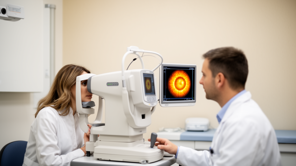
The study, published in BMC Ophthalmology, included 92 participants: 12 with active thyroid orbitopathy, 40 with inactive disease, and 40 healthy controls, and analysed right and left eyes separately using swept-source optical coherence tomography of the optic nerve head.
The researchers set out to evaluate lamina cribrosa morphology in thyroid orbitopathy and to test whether curvature parameters, particularly the lamina cribrosa curvature index, were associated with disease activity or with the presence of the condition. They compared lamina cribrosa thickness, depth, curvature width, curvature depth, and the curvature index across the active disease, inactive disease, and control groups. As a secondary analysis, the active and inactive patient groups were combined for a case–control comparison against the controls.
The results were uniformly negative for the structural parameters under investigation. No statistically significant differences among the three groups were observed in lamina cribrosa thickness, depth, curvature width, curvature depth, or the curvature index, for either right or left eyes. The case–control analysis comparing the combined thyroid orbitopathy group with healthy controls likewise showed no significant differences in any lamina cribrosa parameter, with all comparisons yielding p values above 0.05. A direct pairwise comparison between active and inactive disease also found no statistically significant difference in any parameter.
Not every measured variable was identical between the groups. Hertel exophthalmometry readings, which quantify the degree of forward protrusion of the eyes, and intraocular pressure were both significantly higher in the thyroid orbitopathy groups than in controls, with p values below 0.001. By contrast, sex distribution and axial length did not differ significantly among the three groups, suggesting that these basic demographic and ocular dimensions were unlikely to confound the comparison of lamina cribrosa morphology.
The authors conclude that lamina cribrosa curvature and the related structural parameters did not differ significantly between patients with thyroid orbitopathy and healthy individuals. They suggest that, in patients with thyroid orbitopathy who do not have dysthyroid optic neuropathy, the lamina cribrosa curvature index alone may have limited utility as a structural marker of disease presence or clinical activity. Because the study was cross-sectional and excluded patients with dysthyroid optic neuropathy, the findings speak to the orbitopathy population without that sight-threatening complication rather than to the condition as a whole.
Subject of Research: Lamina cribrosa curvature measured by optical coherence tomography in thyroid orbitopathy
Article Title: Assessment of lamina cribrosa curvature in thyroid orbitopathy: a cross-sectional optical coherence tomography study
Article References: Oyur, G., & Arabacı, İ. Ç. (2026). Assessment of lamina cribrosa curvature in thyroid orbitopathy: a cross-sectional optical coherence tomography study. BMC Ophthalmology. https://doi.org/10.1186/s12886-026-05473-6
Image Credits: AI Generated
DOI: 10.1186/s12886-026-05473-6
Keywords: thyroid orbitopathy, lamina cribrosa, optical coherence tomography, curvature index, optic nerve head, cross-sectional study, dysthyroid optic neuropathy, intraocular pressure, exophthalmometry, BMC Ophthalmology, Assessment, lamina
News Source: Ophelia Keating. (October 29, 2026). Optical coherence tomography finds no lamina cribrosa curvature change in thyroid orbitopathy. Scienmag.