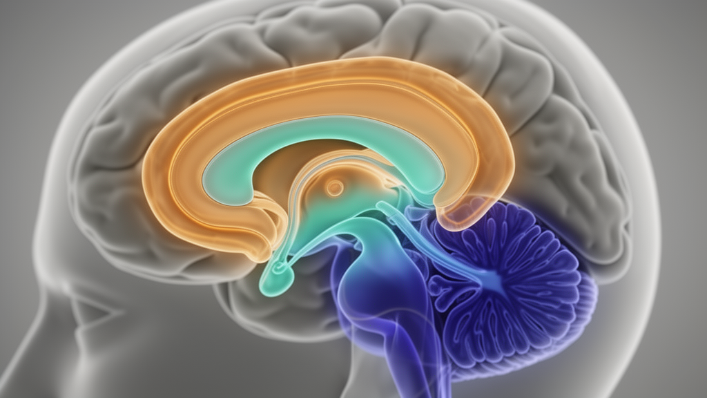
Researchers have established large-scale normative references for hippocampal and hippocampal subfield volumes across the human lifespan, revealing that peak volumes for individual subfields occur at substantially different ages ranging from 27.4 to 40.1 years. Published in Nature Communications, the neuroimaging investigation leveraged high-resolution three-dimensional T1-weighted magnetic resonance imaging scans to characterize structural brain trajectory benchmarks and map anatomical variations across distinct clinical conditions.
To construct the normative volumetric references, the research team analyzed 3D T1-weighted MRI scans acquired from a primary reference cohort of 34,789 healthy individuals spanning an age range of 4 to 85 years. The hippocampus functions as a central hub of cognitive processing, but its internal subfields exhibit varying degrees of vulnerability to the normal aging process. By modeling normative trajectories across this broad lifespan dataset, the authors established baseline volume expectations and quantified the specific developmental and maturational peaks across individual subregions.
Building on these healthy baseline models, the investigators evaluated structural alterations in a clinical population comprising 3,521 patients diagnosed with various neurological disorders. The conditions examined in the study included mild cognitive impairment, Alzheimer’s disease, Parkinson’s disease, cerebral small vessel disease, and multiple sclerosis. For each patient, the researchers derived personalized deviation scores, expressed as centiles relative to the normative reference models, to quantify individualized departures from expected hippocampal and subfield volumes.
The resulting personalized centile scores successfully captured disease-specific patterns of hippocampal subfield abnormality across the five evaluated disorders. Beyond identifying structural deviations, the normative modeling framework supported statistical disease classification, distinguishing between healthy baselines and pathological volume reductions across different neurological diagnoses.
In addition to diagnostic differentiation, the calculated deviation scores demonstrated significant statistical associations with functional patient metrics. Specifically, individual centile scores correlated with measured cognitive performance and tracked longitudinal cognitive outcomes and disease progression over time across the disorders studied.
To facilitate translation into clinical practice, the authors designed a personalized hippocampal health report intended to present individual centile scores and regional volumetric profiles to clinicians. The investigators concluded that normative hippocampal and subfield references provide actionable utility for personalized diagnosis, cognitive assessment, and prognostic evaluation across neurological diseases.
Subject of Research: Normative modeling of hippocampal subfield volumes in neurological disorders
Article Title: Mapping hippocampal subfield heterogeneity across neurological disorders by normative modeling
Article References: Du, X., Gao, P., Chai, L., Zhang, Z., Zhao, X., Wang, W., Huang, P., Lizhu, Y., Wang, P., Wang, Y., Ai, L., Ao, F., Bai, Y., Bai, Y., Bao, H., Cai, Q., Cao, J., Chen, F., Chen, F., … Liu, Y. (2026). Mapping hippocampal subfield heterogeneity across neurological disorders by normative modeling. Nature Communications. https://doi.org/10.1038/s41467-026-79269-z
Image Credits: AI Generated
DOI: 10.1038/s41467-026-79269-z
Keywords: hippocampus, hippocampal subfields, normative modeling, magnetic resonance imaging, structural MRI, T1-weighted MRI, neurological disorders, mild cognitive impairment, Alzheimer's disease, Parkinson's disease, cerebral small vessel disease, multiple sclerosis, centile scores, brain aging, cognitive assessment, disease progression, personalized medicine, neuroimaging
News Source: Cassandra Pierce. (November 11, 2026). Normative Models Map Hippocampal Subfield Variations Across Five Neurological Disorders. Scienmag.