• HOME
  • NEWS
  • EXPLORE
    • CAREER
      • Companies
      • Jobs
    • EVENTS
    • iGEM
      • News
      • Team
    • PHOTOS
    • VIDEO
    • WIKI
  • BLOG
  • COMMUNITY
    • FACEBOOK
    • INSTAGRAM
    • TWITTER
  • CONTACT US
Wednesday, September 27, 2023
BIOENGINEER.ORG
No Result
View All Result
  • Login
  • HOME
  • NEWS
  • EXPLORE
    • CAREER
      • Companies
      • Jobs
        • Lecturer
        • PhD Studentship
        • Postdoc
        • Research Assistant
    • EVENTS
    • iGEM
      • News
      • Team
    • PHOTOS
    • VIDEO
    • WIKI
  • BLOG
  • COMMUNITY
    • FACEBOOK
    • INSTAGRAM
    • TWITTER
  • CONTACT US
No Result
View All Result
Bioengineer.org
No Result
View All Result
Home NEWS Science News Biology

New nanoparticle design paves way for improved detection of tumors

Bioengineer by Bioengineer
June 2, 2021
in Biology
Reading Time: 3 mins read
0
Share on FacebookShare on TwitterShare on LinkedinShare on RedditShare on Telegram

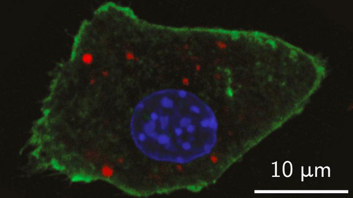
IMAGE

Credit: Giovanni Marco Saladino

Nano-sized particles have been engineered in a new way to improve detection of tumors within the body and in biopsy tissue, a research team in Sweden reports. The advance could enable identifying early stage tumors with lower doses of radiation.

In order to enhance visual contrast of living tissues, state-of-the-art imaging relies on agents such as fluorescent dyes and biomolecules. Advances in nanoparticle research have expanded the array of promising contrast agents for more targeted diagnostics, and now a research team from KTH Royal Institute of Technology has raised the bar further yet. They are combining optical and X-ray fluorescence contrast agents into a single enhancer for both modes.

Muhammet Toprak, Professor of Materials Chemistry at KTH, says the synthesis of contrast agents introduces a new dimension in the field of X-ray bio-imaging. The research was reported in the American Chemical Society journal, ACS Nano.

“This unique design of nanoparticles paves the way for in vivo tumor diagnostics, using X-ray fluorescence computed tomography (XFCT),” Toprak says.

He says the new “core-shell nanoparticles” may have a role to play in the development of theranostics, a portmanteau for therapy and diagnostics, in which for example single drug-loaded particles could both detect and treat malignant tissues.

The core-shell contrast agent gets its name from its architecture: it consists of a core combination of nanoparticles with previously-established potential in X-ray fluorescence imaging, such as ruthenium and molybdenum (IV) oxide. This core is encased in a shell comprised of silica and Cy5.5, a near-infrared fluorescence-emitting dye for optical imaging techniques such as optical microscopy and spectroscopy.

Toprak says that encapsulating the Cy5.5 dye within the silica shell improves the agent’s brightness and extends its photo-stability–enabling the dual optical/X-Ray imaging approach. In addition, silica provides the benefit of tempering the toxic effects of the core nanoparticles.

Tests with laboratory mice have shown that the XFCT contrast agents enable location of early stage tumors of only a few millimetres in size.

Toprak says the technology opens the possibility to identify early stage tumors in living tissue. That’s because the presence of multiple contrast agents increases the odds that diseased areas will show up in scans, even as the distribution of the nanoparticles becomes obscured by their interaction with proteins or other biological molecules.

“Nanoparticles of different size, originating from the same material, don’t appear to be distributed in the blood in the same concentrations,” Toprak says. “That’s because when they come into contact with your body, they’re quickly wrapped in various biological molecules–which gives them a new identity.”

A multitude of contrast agents for XFCT would enable studying the biodistribution of nanoparticles in-vivo using low-dose X-rays, he says. That would allow identifying the best size and surface chemistry of the nanoparticles for the desired targeting and imaging of the diseased region.

###

In addition to Toprak, the study was co-authored by Giovanni M. Saladino, Carmen Vogt, Yuyang Li, Kian Shaker, Bertha Brodin, Martin Svenda and Hans M. Hertz.

Media Contact
David Callahan
[email protected]

Related Journal Article

http://dx.doi.org/10.1021/acsnano.0c10127

Tags: BiotechnologyDiagnosticsHealth CareMedicine/HealthNanotechnology/MicromachinesOptics
Share12Tweet8Share2ShareShareShare2

Related Posts

Figure 4

Living donor liver transplant access is optimal for high-risk waitlisted cirrhosis patients

September 26, 2023
New coronavirus capture material

New material captures coronavirus particles and could transform face mask efficiency

September 26, 2023

UH researcher on team developing sense-and-respond cancer implant technology

September 26, 2023

Invertebrate decline reduces natural pest control and decomposition of organic matter

September 26, 2023
Please login to join discussion

POPULAR NEWS

  • blank

    Microbe Computers

    59 shares
    Share 24 Tweet 15
  • A pioneering study from Politecnico di Milano sheds light on one of the still poorly understood aspects of cancer

    35 shares
    Share 14 Tweet 9
  • Fossil spines reveal deep sea’s past

    34 shares
    Share 14 Tweet 9
  • Scientists go ‘back to the future,’ create flies with ancient genes to study evolution

    75 shares
    Share 30 Tweet 19

About

We bring you the latest biotechnology news from best research centers and universities around the world. Check our website.

Follow us

Recent News

ETRI confirms possibility of wireless communication 40m underground in mine

A novel role discovered for vagus nerve

Patients who quit smoking after percutaneous coronary intervention do as well as non-smokers – unless they had smoked heavily

Subscribe to Blog via Email

Enter your email address to subscribe to this blog and receive notifications of new posts by email.

Join 56 other subscribers
  • Contact Us

Bioengineer.org © Copyright 2023 All Rights Reserved.

No Result
View All Result
  • Homepages
    • Home Page 1
    • Home Page 2
  • News
  • National
  • Business
  • Health
  • Lifestyle
  • Science

Bioengineer.org © Copyright 2023 All Rights Reserved.

Welcome Back!

Login to your account below

Forgotten Password?

Retrieve your password

Please enter your username or email address to reset your password.

Log In