


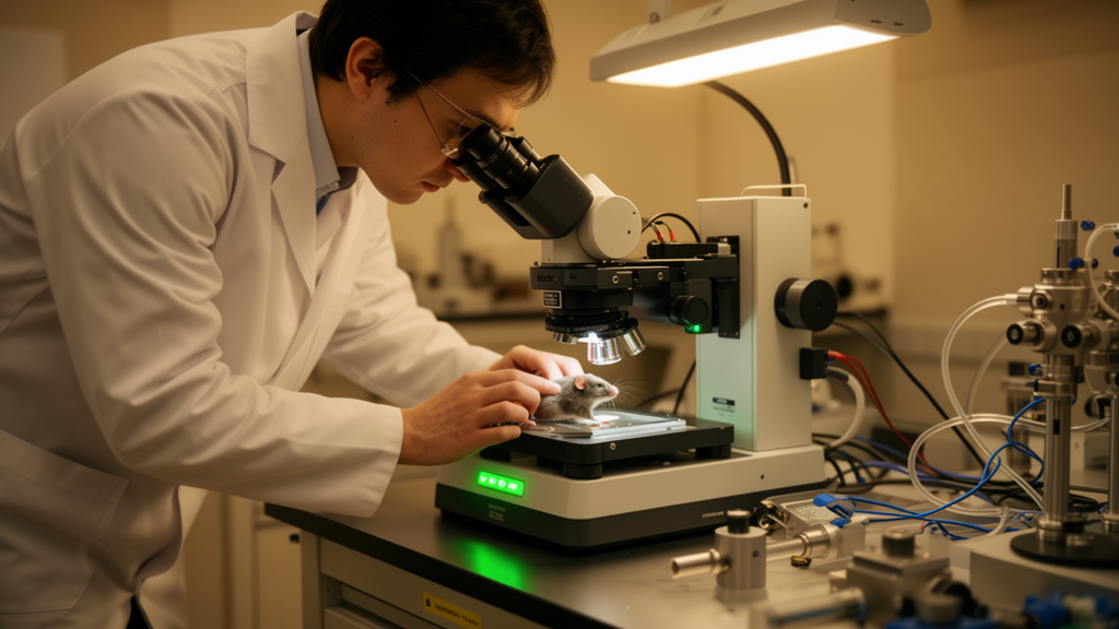
The system, called TrUDI for three-photon ultra-deep imaging, is described in a study published in Nature Methods on 28 October 2026.
The challenge the team set out to address is a fundamental one. Three-photon microscopy has already enabled imaging of neuroactivity at depths of up to roughly 1.2 millimeters in the mouse brain, but the theoretical depth limit of the technique, set by the point at which out-of-focus background fluorescence overwhelms the in-focus signal, lies well beyond 2 millimeters. According to the authors, progress toward that limit has been hampered by insufficient in-focus fluorescence signal, an obstacle they describe as arising from the exponentially hard nature of extending multiphoton imaging depth in scattering brain tissue.
TrUDI tackles this problem through systematic optimization of the in-focus fluorescence signal rather than through a single hardware trick. The approach matches the parameters of the excitation laser, the optical system, and the acquisition process to linear and nonlinear limits for safe laser exposure, while still satisfying the spatiotemporal requirements of single-cell-resolution calcium imaging. In other words, the design extracts as much usable signal as possible without pushing laser exposure beyond safe thresholds.
The payoff is substantial. The researchers report that TrUDI achieves an approximately 250-fold increase in detected fluorescence photon yield at the same laser power as previous approaches. That gain in signal is what enables in vivo cellular-resolution recordings of neuronal population activity within fields of view of roughly 230 by 230 square micrometers at 8.6 hertz, at tissue depths of up to about 2 millimeters in mouse cortex and hippocampus.
The study demonstrates the system’s reach across multiple brain regions and indicator types. In mice expressing the green fluorescent protein Thy1-EGFP, TrUDI enabled deep structural imaging of the hippocampus through the intact posterior parietal cortex. For functional imaging, the system supported calcium recordings over a depth range of approximately 1,800 micrometers, allowing imaging of the majority of hippocampal layers through intact posterior parietal cortex in mice expressing the calcium indicators GCaMP8m and GCaMP8s.
The depth advantage extends beyond the hippocampus. In mice expressing the indicator GCaMP6s, TrUDI enabled deep-tissue calcium imaging over a depth range of approximately 2,000 micrometers within motor cortical areas and the striatum.
The TrUDI design addresses this through targeted excitation, which the authors report minimizes laser exposure within the system. By concentrating excitation where signal is needed and matching exposure to established safety limits, the approach aims to keep deep imaging compatible with live-animal experiments.
The work is a methods development study conducted in mice, and its reported capabilities reflect the specific indicators, brain regions, and imaging parameters described in the paper. The abstract frames the contribution as approaching the fundamental depth limit of three-photon imaging, which is determined by the depth at which background fluorescence exceeds in-focus signal, rather than as surpassing that limit.
Subject of Research: Deep three-photon microscopy of neuronal population activity in the mouse brain
Article Title: Cellular-resolution three-photon ultra-deep imaging (TrUDI) of neuronal population activity at 2-mm depth in the mammalian brain
Article References: Santra, K., Barber, K., Logiacco, F., Frechou, M. A., Bellafard, A., Liou, J.-Y., & Vaziri, A. (2026). Cellular-resolution three-photon ultra-deep imaging (TrUDI) of neuronal population activity at 2-mm depth in the mammalian brain. Nature Methods. https://doi.org/10.1038/s41592-026-03251-2
Image Credits: AI Generated
DOI: 10.1038/s41592-026-03251-2
Keywords: three-photon microscopy, TrUDI, neuronal imaging, calcium imaging, hippocampus, mouse brain, deep-tissue imaging, neuroscience, fluorescence, laser exposure, Nature Methods, brain imaging
News Source: Cassandra Pierce. (October 28, 2026). New microscopy method reaches 2-mm-deep neuronal imaging in the mouse brain. Scienmag.



