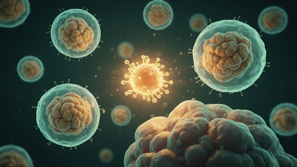
Oncolytic viruses (OVs) have emerged as one of the most intriguing strategies in modern cancer immunotherapy, capable of selectively infecting and destroying tumor cells while sparing healthy tissue. Yet a new review published in Experimental Biology and Medicine suggests that the success of this approach may hinge on an unexpected player: neutrophils, the most abundant white blood cells in the human body. The review, led by researchers at Foshan University in China, systematically examines how these cells can either amplify or undermine the therapeutic effects of oncolytic virotherapy, offering a detailed roadmap for optimizing clinical applications.
The concept of using viruses to fight cancer is not new. Reports dating back to 1904 described unexpected remission of leukemia symptoms in a patient following influenza infection, and in 1912, Italian physician Depace documented spontaneous regression of cervical cancer after administration of an attenuated rabies vaccine. These early observations spurred interest in the potential oncolytic properties of viruses, though initial attempts to inject live wild-type viruses into cancer patients often resulted in significant toxicity. The field evolved through three distinct stages: direct use of wild-type viruses, targeted genetic modification of viral genomes in the 1990s to enable tumor-specific replication, and, in the current century, insertion of exogenous genes to enhance therapeutic effects. In 2015, Talimogene laherparepvec became the first OV therapy to receive regulatory approval worldwide, and since then, an increasing number of OV candidates have entered clinical trials.
Oncolytic viruses exert their antitumor effects through three primary mechanisms. First, they directly lyse tumor cells by selectively replicating within them. Teserpaturev (G47Δ), an OV approved for clinical use in Japan, targets aberrant signaling pathways in gliomas to induce tumor cell death. Second, OVs activate the host immune system. As pathogen-associated molecular patterns, they stimulate innate immune cells including macrophages and natural killer cells, while engineered OVs expressing immunomodulatory genes such as GM-CSF can promote T cell activation and trigger adaptive immune responses. Third, OVs can disrupt tumor vasculature. The NDV-GT OV, for instance, induces expression of the porcine α1,3GT gene on tumor cell surfaces, generating α-Gal antigens that lead to tumor vascular embolism and necrosis, with a disease control rate of 90% in clinical trials.
Neutrophils originate from the bone marrow and account for approximately 50% to 70% of total leukocytes in mammalian blood. Within the tumor microenvironment (TME), these cells can polarize into two distinct phenotypes: N1 and N2. N1 neutrophils typically appear during early stages of tumor development and primarily function to inhibit tumor growth, exhibiting antitumor properties and enhancing cytotoxic T lymphocyte activity. In contrast, N2 neutrophils are more prevalent in later stages and predominantly promote tumor progression. Transforming growth factor-beta (TGF-β) regulates the transition of neutrophils toward the N2 phenotype. In a pancreatic cancer liver metastasis model, TGF-β secreted by tumors activates SMAD3 signaling in neutrophils, inducing expression of nuclear factor erythroid 2 and subsequently activating peptidyl-arginine deiminase 4, driving polarization toward the N2 phenotype and promoting tumor metastasis.
The review highlights several mechanisms through which neutrophils positively regulate oncolytic virotherapy. Their inherent chemotactic and tumor-homing abilities allow them to carry OVs across physiological barriers, including the blood-brain barrier, achieving targeted delivery. After intravenous injection, OVs can be captured by receptors on neutrophil surfaces, forming a virus-neutrophil complex. In a clinical trial, investigators observed that increased intratumoral neutrophil infiltration in gastric cancer patients with OV therapy was associated with exacerbated CD8+ T cell exhaustion. Inspired by this observation, they designed a bifunctional bioorthogonal delivery system using an anti-Ly6G antibody modified with DBCO to tag circulating neutrophils, significantly boosting virus load inside the tumor. Once the virus reached the tumor site, the neutrophils underwent apoptosis, relieving immunosuppression and reducing T cell exhaustion, effectively turning neutrophils from a therapeutic obstacle into an efficient delivery vehicle.
Beyond delivery, neutrophils can directly enhance tumor killing. In a mouse model of lung melanoma metastasis, treatment with orf virus significantly increased the number of neutrophils in peripheral blood, peritoneal cavity, spleen, liver, and the TME. These ORFV-activated neutrophils directly killed tumor cells through secretion of TNF-α and expressed major histocompatibility complex class II, contributing to immunomodulatory functions within the TME. Similarly, vesicular stomatitis virus (VSV) infection triggers rapid release of neutrophils from the bone marrow, which transition from a mature to an immature phenotype and exhibit enhanced antigen-presenting capacity and elevated secretion of inflammatory cytokines, promoting T cell-mediated antitumor immune responses.
However, neutrophils can also negatively regulate OV therapy through their antiviral activity. As the first line of defense in the immune system, they secrete various antimicrobial and antiviral substances, including myeloperoxidase, defensins, and antimicrobial peptides. Most oncolytic vaccinia viruses are phagocytosed and degraded by neutrophils in the bloodstream. Depletion of neutrophils using anti-Ly6G antibodies leads to increased accumulation of circulating oncolytic vaccinia viruses in peripheral blood and enhanced viral deposition at tumor sites, potentiating antitumor efficacy. Neutrophils can also suppress OV activity through the release of neutrophil extracellular traps (NETs), structures composed of genomic DNA, histones, defense proteins, and proteases that trap and inactivate viruses. In oHSV therapy, upregulation of insulin-like growth factor 2 mRNA-binding protein induces NET formation, thereby reducing oncolytic activity.
Furthermore, neutrophils in the TME can mediate an immunosuppressive microenvironment that undermines OV efficacy. They induce the expansion of regulatory T cells (Tregs), which suppress the activity of cytotoxic T cells and natural killer cells by secreting inhibitory cytokines such as interleukin-10 and TGF-β. Neutrophils also interact with myeloid-derived suppressor cells (MDSCs), facilitating their maturation and functional competence. MDSCs accumulate extensively in the TME and inhibit T cell proliferation and activation by secreting arginase I, nitric oxide, and reactive oxygen species, representing one of the core mechanisms by which tumors achieve immune escape.
The review authors argue that the future of OV optimization should not rely on blunt depletion of the entire neutrophil compartment, which would clinically recapitulate the fatal condition of leukocyte adhesion deficiency and predispose patients to life-threatening infections. Instead, they advocate for more refined and precise immunomodulatory approaches. For example, targeting specific chemokine receptors such as CXCR2 or selectively interfering with particular immunosuppressive subsets can block pathological recruitment while preserving normal immune surveillance. In a phase II clinical trial, the CXCR2 antagonist navarixin in combination with pembrolizumab for advanced solid tumors induced neutropenia without associated fever or infection. Similarly, the selective PAD4 inhibitor JBI-589 transcriptionally downregulates CXCR2 expression, precisely blocking neutrophil migration into the TME and enhancing the efficacy of immune checkpoint inhibitors.
This conceptual shift from global depletion to precision modulation may represent the key to advancing OV-based combination immunotherapy toward safe and effective clinical application. The authors emphasize that future translational research should focus on temporally dynamic regulation of neutrophil functions, spatial blockade of pathological recruitment to tumor sites, synergistic combination with immune checkpoint inhibitors, and clinically precise patient stratification based on neutrophil-related biomarkers. Given the complexity and heterogeneity of the TME, neutrophil responses vary from patient to patient, making personalized treatment strategies essential for predicting and improving therapeutic outcomes in oncolytic virotherapy.
Subject of Research: Immunology
Article Title: The role and mechanism of neutrophils in oncolytic virus therapy
Article References: Huang, B., Yang, Q., Wang, R., & Zhao, C. (2026). The role and mechanism of neutrophils in oncolytic virus therapy. Experimental Biology and Medicine, 251, Article 11192. https://doi.org/10.3389/ebm.2026.11192
Image Credits: AI Generated
Keywords: oncolytic viruses, neutrophils, cancer immunotherapy, tumor microenvironment, N1/N2 polarization, virotherapy, role, mechanism, oncolytic, virus, therapy, scientific research
News Source: Kristina Jarvis. (October 17, 2026). Neutrophils: The Double-Edged Immune Cells Shaping Oncolytic Virus Therapy. Scienmag.