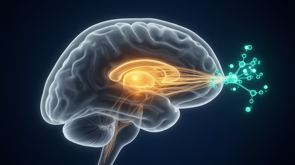
A neonatal injury to a small but influential brain structure can leave lasting marks on how the brain handles attention, according to a new rat study published in Frontiers in Behavioral Neuroscience. Researchers report that damaging the mediodorsal thalamus (MD) in newborn rats produced a persistent impairment of latent inhibition, a form of selective attention, alongside elevated dopamine D2 receptor expression in the prefrontal cortex and reduced recruitment of the prefrontal cortex and nucleus accumbens during learning. Notably, a single well-known drug intervention during a critical phase of the task restored both the behavioral deficit and the abnormal neuronal activation patterns.
The mediodorsal thalamus is a higher-order thalamic nucleus with a central role in the maturation and function of prefrontal cortical circuits. Developmental dysfunction of this region has been implicated in the cognitive deficits associated with neurodevelopmental disorders, but the molecular and circuit-level mechanisms behind that link have remained poorly understood.
When the rats reached adulthood, the researchers assessed latent inhibition using a conditioned taste aversion paradigm. The MD-lesioned animals showed a persistent impairment of this effect, meaning their brains processed pre-exposed stimuli differently than healthy controls did.
To understand what was happening inside the circuit, the team measured neuronal activation using c-Fos immunohistochemistry, a technique that marks neurons that were active during a specific task. This suggests that early thalamic damage disrupts the coordinated recruitment of the MD–PFC–NAc circuit during selective attention tasks in adulthood.
The functional changes were accompanied by molecular remodeling. Using immunohistochemistry, Western blotting, and quantitative PCR, the researchers found a selective increase in prefrontal D2 receptor expression at the transcript, protein, and cellular levels. Importantly, this increase was specific to the prefrontal cortex: no comparable changes were observed in the secondary motor cortex, indicating that the effect was not a general response to the lesion but a targeted alteration of dopaminergic signaling in the prefrontal circuit.
The most striking result came when the researchers administered haloperidol, a dopamine D2 receptor antagonist, during the stimulus pre-exposure phase of the latent inhibition task. In the MD-lesioned animals, haloperidol restored latent inhibition and normalized c-Fos activation in both the prefrontal cortex and the nucleus accumbens. This pattern suggests that the behavioral deficit in the lesioned animals was linked to the elevated D2 receptor signaling, and that temporarily dampening that signaling during learning could compensate for it.
The authors interpret these findings as evidence that early MD dysfunction induces long-lasting remodeling of prefrontal dopaminergic signaling and disrupts the recruitment of the MD–PFC–NAc circuit during selective attention.
As with any animal study, the findings come with important caveats. The work was conducted in rats with experimentally induced lesions on postnatal day 4, a model of developmental disruption rather than a naturally occurring condition, and the results were measured in adulthood after the brain had developed around the injury. The haloperidol effect was specific to the timing of administration during stimulus pre-exposure and to the lesioned animals, and the study does not establish that the same intervention would be effective or appropriate in humans. Translating these circuit-level mechanisms to clinical contexts will require further research.
Nevertheless, the study offers a concrete example of how a developmental insult to one brain region can cascade into molecular and circuit-wide changes elsewhere, and how those changes can be traced to a specific receptor system. By linking a neonatal thalamic lesion to altered prefrontal D2 receptor expression, reduced prefrontal and accumbal activation during learning, and a pharmacologically reversible attentional deficit, the work provides a framework for understanding how early thalamic dysfunction might contribute to lasting cognitive impairments, and points to dopamine D2 signaling as a potential target for future investigation.
Subject of Research: Effects of neonatal mediodorsal thalamus lesions on prefrontal dopamine D2 receptors and latent inhibition in rats
Article Title: Developmental disruption of the mediodorsal thalamus alters prefrontal dopamine D2 receptor expression and prefrontal–accumbal neuronal recruitment during latent inhibition
Article References: Ouhaz, Z., Iben-Daoudi, H., Darfaoui, M., Tait, D. S., Lamghari Moubarrad, F.-Z., Ba-M’Hamed, S., Nejmeddine, M., & Bennis, M. (2026). Developmental disruption of the mediodorsal thalamus alters prefrontal dopamine D2 receptor expression and prefrontal–accumbal neuronal recruitment during latent inhibition. Frontiers in Behavioral Neuroscience, 20, 1998199. https://doi.org/10.3389/fnbeh.2026.1998199
Image Credits: AI Generated
DOI: 10.3389/fnbeh.2026.1998199
Keywords: mediodorsal thalamus, latent inhibition, dopamine D2 receptor, prefrontal cortex, nucleus accumbens, haloperidol, c-Fos, neurodevelopment, rats, selective attention, conditioned taste aversion, Developmental
News Source: Harold Sullivan. (October 29, 2026). Neonatal Thalamus Disruption Rewires Prefrontal Dopamine Signaling in Rats. Scienmag.