
In a groundbreaking study conducted by a team of researchers at Kindai University, a novel understanding of neuronal migration has been unveiled, shedding light on the dynamic processes that govern how neurons navigate the complexities of the developing brain. Neurons are not merely passive cells drifted by the flow of biological currents; they actively employ sophisticated strategies to reach their final destinations within the organism while responding to fluctuating environmental conditions. This adaptability is vital for the proper formation and function of neural networks, making the study’s findings particularly significant in the fields of developmental biology and neuroscience.
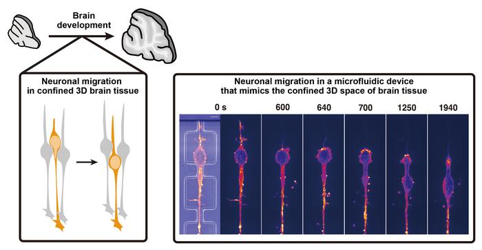
Employing cutting-edge microfluidic technology, the research team led by Dr. Naotaka Nakazawa observed that neuronal movement is far more intricate than previously understood. The study offers insights into how neurons adjust their migration strategies based on the surrounding environment—specifically, whether they are traversing a flat, unencumbered surface or squeezing through confined, three-dimensional spaces typical of brain tissue. In simpler terms, neurons adapt their locomotion methods depending on their spatial constraints, much like a person would alter their walking style when moving through a crowded space versus an open area.
Key to this transformation in migration tactics is the role of the protein PIEZO1, which serves as a mechanosensitive channel that detects mechanical forces acting on the neurons. As these cells face physical constraints, PIEZO1 becomes activated, prompting an influx of calcium ions into the cytosol. This calcium influx triggers a signaling cascade within the neuron that restructures its internal architecture, reallocating motor proteins that drive movement. In less confined environments, these proteins are concentrated at the front of the neuron, facilitating a pulling motion that propels cellular movement. Conversely, in tight spaces, the reorganization pushes these motor proteins toward the rear of the cell, generating the necessary force to navigate through constricted areas.
The implications of this research extend beyond mere academic interest; they hold the potential to influence therapeutic strategies for neurological conditions. Damage to the brain can severely impair neuronal function and survival. By enhancing our understanding of how neurons migrate in response to their environment, there may be innovative avenues for promoting neural repair and regeneration in response to brain injuries. Research indicates that neuroblasts, or neuronal precursors, migrate toward lesions to facilitate recovery. Insights from this study could inform approaches aimed at enhancing this natural repair mechanism and restoring functional capacity in affected brains.
Furthermore, the adaptability demonstrated by neurons in the study raises critical questions about the biological underpinnings of cell migration across various contexts. Migration is a fundamental process not confined to the central nervous system; it also plays pivotal roles in embryonic development, immune responses, and cancer metastasis. The findings suggest that similar strategies employed by neurons may also be harnessed by cancer cells as they traverse and invade different tissue environments. Grasping the mechanics of cellular movement in response to physical constraints could revolutionize diagnostic and therapeutic protocols across an array of medical paradigms.
The research prominently highlights distinct migration strategies observed among different neuron types. For instance, forebrain interneurons utilize myosin to exert force on their nucleus, which contrasts sharply with the actomyosin-fueled pulling motion witnessed in cerebellar granule neurons cultured in standard laboratory dishes. Despite these differences in mechanisms, the findings posit that the ability to switch between these strategies might not strictly be determined by the neuron type but rather by the immediate physical environment, fostering a paradigm shift in our comprehension of neuronal behavior.
The study also delves into the morphological changes neuronal nuclei undergo during migration. Nakazawa’s team elucidated how migrating neurons frequently exhibit significant deformation of their nuclei, particularly when passing through confined environments. This deformation reflects the physical stresses encountered within the tissue and emphasizes the dynamic nature of cellular morphology as cells respond to their surroundings. Such insights warrant further investigation into the biomechanical properties of neurons, unveiling a captivating intersection between form and function in cellular behavior.
This pioneering research paves the way for subsequent studies challenging entrenched notions of neuronal migration and could propel further inquiries into the relationship between mechanical properties and cellular signaling pathways. The nuances of how cells sense and respond to their environments remain an exciting frontier in biology. Future work could explore the broader applicability of these findings across various cell types and organisms, leading to a deeper comprehension of how life forms navigate their complex habitats.
In addition to its scientific implications, this research stresses the importance of interdisciplinary collaboration in understanding complex biological phenomena. The collaboration between scientists from various institutions, including Kyoto University and the National University of Singapore, exemplifies how diverse expertise can lead to groundbreaking discoveries. Such partnerships foster a rich ecosystem for scientific inquiry, driving forward the frontiers of knowledge and understanding in significant ways.
In summary, the paradigm established by Dr. Nakazawa and his research team represents a crucial step in our understanding of neuronal migration, emphasizing the role of environmental mechanics in guiding cell movement. As our grasp of these complex processes evolves, so too will our ability to devise innovative therapies for a host of neurological conditions, ultimately aiming to enhance brain health and function.
With each new revelation about how neurons adapt to their environment, the scientific community edges closer to deciphering the intricate behaviors underlying cell migration. As researchers continue to ponder questions left unanswered by this study, the future of neurobiology promises to be characterized by awe-inspiring discoveries that can reshape our comprehension of both development and pathology within the realm of the nervous system.
Subject of Research: Animals
Article Title: PIEZO1-dependent mode switch of neuronal migration in heterogeneous microenvironments in the developing brain
News Publication Date: 25-Mar-2025
Web References: DOI Link
References: Not available
Image Credits: Naotaka Nakazawa, Kindai University, Japan
Keywords: Neurons, Brain development, Neuronal migration, PIEZO1, Developmental neuroscience, Mechanotransduction, Cell biology, Experimental study
Tags: adaptability of neuronsbrain tissue navigation challengesconfined spaces and neuronal locomotiondevelopmental neuroscience insightsdynamic processes in brain developmentenvironmental influences on neuron movementKindai University research findingsmicrofluidic technology in researchnavigating dense brain tissueneural network formationneuronal migration strategiesPIEZO1 protein function
