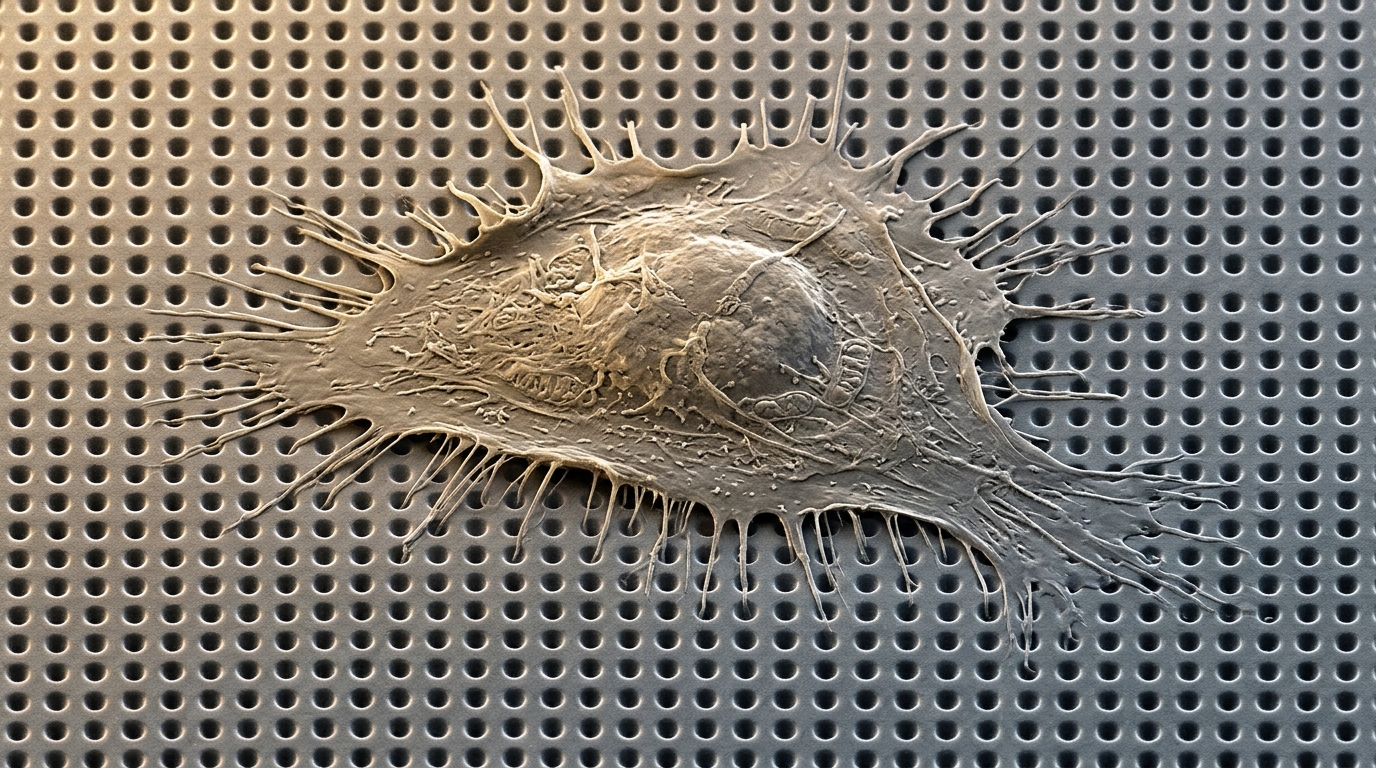
Nanopit arrays significantly enhance the spreading area and early osteogenic differentiation of human adipose-derived stem cells compared with inverted nanopillar arrays. Investigating how nanotopographic architectures guide stem cell behavior, researchers evaluated cellular morphological adjustments, cell viability, and early bone-lineage commitment on nanoscale patterns. The study revealed that nanoscale pits provide continuous adhesive pathways that facilitate wider cellular expansion and robust focal adhesion development, yielding elevated expression of runt-related transcription factor 2, a key early osteogenic gene marker. In contrast, isolated nanopillar arrays limited cell spreading and focal adhesion formation, demonstrating that spatial continuity across synthetic surfaces critically regulates mechanosensitive cellular responses.
Fabrication of the nanostructured substrates relied on colloidal lithography and capillary force lithography to yield precise inverted nanotopographies of nanopillars and nanopits across three defined pitches. Quartz master molds were initially produced using polystyrene nanobeads having diameters of 350 nanometers, 500 nanometers, and 1000 nanometers to regulate center-to-center feature spacing. Droplets of polyurethane acrylate precursor were dispensed onto the master molds, covered with glass coverslips, filled through capillary force, cured under 300 millijoules per square centimeter of ultraviolet exposure, and peeled. Polyurethane acrylate nanopit arrays served as secondary molds with anti-adhesion coatings to replicate corresponding nanopillar arrays. The resulting nanopillars exhibited mean diameters of 208 ± 9 nanometers with 142-nanometer nanogaps at 350-nanometer pitch, 292 ± 12 nanometers with 208-nanometer nanogaps at 500-nanometer pitch, and 623 ± 23 nanometers with 377-nanometer nanogaps at 1000-nanometer pitch. Corresponding nanopits exhibited mean diameters of 253 ± 11 nanometers, 359 ± 12 nanometers, and 787 ± 19 nanometers, with all fabricated surfaces uniformly coated with fibronectin for integrin-mediated cellular attachment.
Cellular assays utilized human adipose-derived mesenchymal stem cells obtained from commercial repositories at their sixth passage to assess adhesion dynamics and differentiation potential. Cells were seeded onto the fibronectin-coated nanostructured substrates in twelve-well plates at an initial density of 20,000 cells per well. Primary cell maintenance was performed in low-glucose Dulbecco’s modified Eagle’s medium basal media maintained within a humidified incubator at 37 degrees Celsius with 5 percent carbon dioxide. Following an initial 24-hour attachment period, cultures were washed with phosphate-buffered saline to eliminate unattached cells before proceeding with subsequent phenotypic evaluations. For osteogenic differentiation investigations, the basal culture medium was replaced after 24 hours of attachment with specialized osteogenic differentiation medium. Cells were then continuously cultured for 14 days under standard incubation conditions, receiving routine medium exchanges every three to four days to sustain metabolic stability and lineage induction throughout the two-week developmental timeline.
Evaluating cellular tolerance to the synthetic substrates, the researchers quantified metabolic viability using the colorimetric 3-(4,5-dimethyl-thiazol-2-yl)-2,5-diphenyltetrazolium bromide assay after 24 hours of continuous cell-substrate interaction. Subsequently, two hundred microliters of dimethyl sulfoxide was introduced per well to thoroughly dissolve the accumulated formazan precipitate. Solution absorbance was measured at 570 nanometers using a VersaMax microplate reader across six independent experimental wells per condition.
High-resolution scanning electron microscopy was conducted to characterize cell morphology and spreading behavior across the various nanostructured surfaces following 24 hours of cultivation. Adherent cells were initially fixed using Karnovsky’s fixative containing 2 percent glutaraldehyde and 2 percent paraformaldehyde before undergoing dehydration through a graded ethanol series spanning 50, 70, 80, 90, and 100 percent concentrations. Following critical-point drying, a thin conductive gold layer was deposited on the specimens via sputtering in a cluster system, allowing visualization under a JEOL JSM-7100F field-emission scanning electron microscope. Calibrated 40-times magnification micrographs with a resolution of 0.46 pixels per micrometer were analyzed using ImageJ software. Cell boundaries were traced to measure projected cell area, aspect ratio, major axis length, and minor axis length across 35 individual cells per experimental condition, with statistical comparisons performed using one-way analysis of variance and post-hoc testing at a significance threshold of P < 0.05.
Scanning electron microscopy demonstrated that periodic nanostructures provided effective contact guidance, causing stem cells to align their spreading boundaries with the underlying structural symmetry relative to unpatterned controls. Cells cultured on 350-nanometer and 500-nanometer pitches spread extensively, displaying characteristic triangular protrusions at spreading edges, whereas cells grown on 1000-nanometer pitches assumed elongated, slender configurations. Across all evaluated dimensions, nanopit arrays yielded significantly larger total cell areas than corresponding nanopillar arrays. On nanopit arrays, filopodia extended along connecting ridges and spanned across individual pits when walls were narrow. Conversely, on nanopillar arrays, filopodia aligned along symmetrical pillar axes. At the largest 1000-nanometer pitch, wider gaps of 377 nanometers between pillars and 787-nanometer pit openings inhibited lateral cell spreading, causing a pronounced reduction in minor cell axis length. This transverse contraction generated significantly elevated aspect ratios for both topographies, highlighting how nanoscale feature spacing constrains cell elongation and spreading symmetry.
Focal adhesion formation and actin cytoskeleton organization were investigated after 24 hours of incubation using multi-channel immunofluorescent confocal microscopy. Adherent stem cells were fixed with 4 percent paraformaldehyde in phosphate-buffered saline, permeabilized with 0.5 percent Triton X-100, and blocked with 5 percent bovine serum albumin. Samples were incubated with primary antibodies against vinculin and Alexa Fluor 633-conjugated phalloidin, followed by Alexa Fluor 488-labeled secondary antibodies and 4′,6-diamidino-2-phenylindole nuclear counterstaining. Fluorescence micrographs were acquired at 40-times magnification using a Carl Zeiss LSM700 confocal microscope and analyzed through ImageJ following established quantitative protocols across five individual cells per group. While total focal adhesion area and adhesion number showed negligible divergence between nanopillars and nanopits at the 1000-nanometer pitch, nanopit arrays supported significantly higher focal adhesion numbers and greater aggregate adhesion area at 350-nanometer and 500-nanometer pitches, confirming that continuous nanostructured interfaces promote robust multi-protein anchoring complexes.
The observed divergence in focal adhesion maturation reflects fundamental physical differences in how cells perceive continuous versus discontinuous adhesive interfaces. On nanopit arrays, neighboring depressions are bounded by interconnected polymeric ridges coated with fibronectin, presenting an uninterrupted planar pathway that facilitates lamellipodial extension and stable integrin clustering. Conversely, nanopillar arrays restrict adhesive interactions to isolated pillar summits separated by empty nanogaps, forcing protruding filopodia to bridge discontinuous gaps. Cross-sectional electron microscopy from related literature confirmed that cell-substrate contact remains predominantly confined to upper feature surfaces rather than recessed voids. Furthermore, connected nanopit arrays offer higher effective structural rigidity than discrete nanopillars, which experience increased lateral compliance and bending at narrow 350-nanometer pitches. As focal adhesion assembly couples directly to intracellular actin stress fibers, the enhanced adhesion maturation on nanopits generates greater cellular traction forces, providing a biomechanical foundation that favors osteogenic lineage commitment over compliant, interrupted topographies.
Differentiation outcomes were evaluated after 14 days of osteogenic induction by measuring transcriptional levels of runt-related transcription factor 2 via quantitative reverse transcription polymerase chain reaction. Total cellular ribonucleic acid was extracted using commercial silica-column purification kits, and 500 nanograms of purified RNA was reverse-transcribed into complementary deoxyribonucleic acid under thermal cycling conditions of 42 degrees Celsius for 60 minutes and 94 degrees Celsius for 5 minutes. Quantitative amplification was performed using targeted primers, with glyceraldehyde-3-phosphate dehydrogenase serving as the endogenous housekeeping reference for comparative cycle threshold analysis across duplicate biological replicates. Experimental data demonstrated that RUNX2 expression was markedly elevated in stem cells cultured on nanopit arrays compared to nanopillar arrays across matching pitches. Notably, the relative transcriptional pattern of RUNX2 closely mirrored the quantitative hierarchy observed in projected cell spreading area, reinforcing an empirical correlation between planar cell expansion and early osteogenic activation.
While the experimental data demonstrate that nanopit arrays enhance early osteogenesis in human adipose-derived stem cells, the investigators identified several experimental limitations and prospective research requirements. Osteogenic evaluation in this study was restricted to RUNX2 expression at day 14, representing early transcription factor activation prior to extracellular matrix mineralization; assessing late-stage osteogenic markers, such as alkaline phosphatase and osteocalcin, alongside matrix mineralization assays remains necessary to confirm mature bone formation. Additionally, while literature links traction-dependent mechanotransduction to the RhoA-ROCK signaling pathway, the authors did not directly measure RhoA activity or enzymatic ROCK phosphorylation, maintaining that pathway involvement remains a supported hypothesis requiring validation through pharmacological inhibitor assays and traction force microscopy. Future investigations must also incorporate three-dimensional z-resolved confocal imaging, correlative light-electron microscopy to verify focal adhesion localization, systematic evaluations of substrate stiffness and structural disorder, and in vivo animal implant models to advance clinical translation.
Subject of Research: Nanotopographical effects on stem cell morphology and osteogenic differentiation
Article Title: The effects of nanopillar and nanopit arrays on the morphology and osteogenic differentiation of adipose-derived stem cells
Article References: Kang, J., Yun, Y.-S., Kang, E.-H., Lee, J., Jeon, D.-J., Ji, S., Kim, Y.-O., Yun, I.-S., & Yeo, J.-S. (2026). The effects of nanopillar and nanopit arrays on the morphology and osteogenic differentiation of adipose-derived stem cells. Applied Microscopy, 56(1), Article 2. https://doi.org/10.1186/s42649-026-00122-0
Image Credits: AI Generated
DOI: 10.1186/s42649-026-00122-0
Keywords: Nanotechnology, Stem Cells, Osteogenic Differentiation, Nanopillars, Nanopits, Focal Adhesions, Biomimetic Scaffolds, Cell Morphology, effects, nanopillar, nanopit, arrays
News Source: Drew Townsend. (November 6, 2026). Nanopit Arrays Outperform Nanopillars in Promoting Stem Cell Spreading and Osteogenesis. Scienmag.