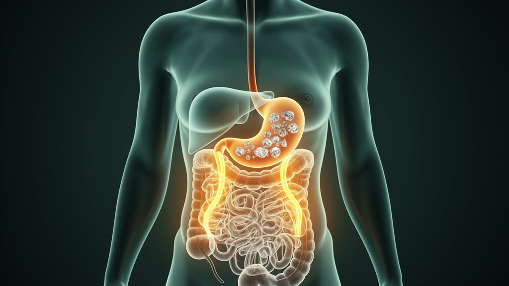
Gallstone disease affects a substantial share of the world’s population, and its prevalence rises steeply in people living with obesity. Yet the molecular events that turn cholesterol suspended in bile into hard, painful stones remain only partly understood. A new study published in the International Journal of Obesity by Jacqueline B. Hoozemans of Amsterdam University Medical Centers and colleagues takes an unusually broad look at the problem, combining gene expression, circulating metabolites, and gut microbial DNA from a well-characterized group of women undergoing bariatric surgery. The results challenge several assumptions about where, in the body, gallstone disease begins.
The team drew on the BARIA cohort, a longitudinal study designed to untangle the relationships between gut microbiota and host metabolism in severe obesity. From this cohort they enrolled 108 consecutive female patients, all with obesity and all scheduled for surgery. At baseline, ultrasound examination revealed gallstones in 39 of these women, who served as cases, while the remaining 69 were stone-free and served as controls. Because the participants were recruited consecutively and matched for the circumstance of surgery, the design reduces some of the confounding that plagues comparisons of gallstone patients drawn from different clinical settings.
What makes the study distinctive is the breadth of tissue sampling. During surgery, the researchers collected biopsy specimens from the liver, from visceral adipose tissue deep in the abdomen, and from subcutaneous adipose tissue beneath the skin. They also collected fasting blood for untargeted plasma metabolomics and stool for fecal shotgun metagenomics, a technique that sequences the genomes of gut microbes directly rather than relying on marker genes. Bulk RNA sequencing was then used to measure gene expression in each tissue type, allowing the investigators to ask which organs showed molecular signatures that distinguished gallstone carriers from stone-free controls.
The clearest signal emerged from visceral adipose tissue. Fifty-seven genes were expressed at different levels in the visceral fat of women with gallstones compared with controls, a pattern that held up in the primary analysis. The liver, by contrast, showed differences in gene expression only after secondary, more granular analysis. Subcutaneous fat, often treated as a relatively inert storage depot in metabolic studies, contributed less to the molecular picture. Taken together, the findings suggest that the fat surrounding the abdominal organs, rather than the liver alone, may be an underappreciated participant in gallstone formation.
Perhaps the most surprising result concerns the classic textbook story of gallstone genetics. Decades of work, including genome-wide association studies that have identified multiple susceptibility loci, have pointed to hepatic ABC transporters, membrane proteins that pump cholesterol and phospholipids into bile, as central players in determining whether bile becomes supersaturated with cholesterol. In this cohort, those classic transporter genes did not differ between cases and controls. Instead, a different cholesterol transporter, ABCG1, emerged, and its expression in the liver was decreased in women with gallstone disease. ABCG1 is known to mediate cholesterol efflux to high-density lipoprotein and to protect cells from lipid accumulation, so reduced expression could plausibly shift cholesterol handling in ways that favor crystallization, though the authors are careful to frame this as an association rather than a demonstrated cause.
The metabolomic data added a second layer of evidence. Plasma from the gallstone group contained increased levels of both primary bile acids, which are synthesized in the liver from cholesterol, and secondary bile acids, which are produced when gut bacteria chemically modify the primary forms. Elevated circulating bile acids in stone formers is consistent with a model in which bile acid supply relative to cholesterol secretion is a key determinant of lithogenic bile; prior work has argued that cholesterol-supersaturated gallbladder bile in gallstone patients preferentially develops from a shortage of bile acids rather than an excess of cholesterol. The persistence of these differences in fasting plasma suggests a systemic metabolic signature rather than a purely local gallbladder phenomenon.
By contrast, the gut microbiome results were strikingly muted. Despite the considerable attention that intestinal microbes have received as modulators of bile acid metabolism, fecal shotgun metagenomics revealed only limited differences in microbial composition between women with gallstones and those without. This finding aligns with some prior reports, including a population study that found carrying asymptomatic gallstones was not associated with altered intestinal microbiota composition or diversity, while cholecystectomy, the surgical removal of the gallbladder, was associated with significant dysbiosis. The new data suggest that, at least in women with obesity who have not yet had their gallbladders removed, the microbiome is not the dominant driver of stone formation.
The study’s focus on women is deliberate and scientifically motivated. Obesity is a well-established causal risk factor for symptomatic gallstone disease, an association supported by Mendelian randomization analyses that use genetic variants to disentangle correlation from causation. But female sex adds its own risk, in part through estrogen signaling pathways that have been shown in mouse models to promote cholesterol crystallization in the gallbladder via two distinct receptors. Sex differences in lipid metabolism are also known to be influenced by the presence of the gut microbiota. By restricting the analysis to adult women with obesity, the investigators removed sex as a confounder, at the cost of limiting generalizability to men.
The work also builds on the group’s own earlier findings. Previous analyses from the same Amsterdam collaboration had shown that gallstone formation follows a different trajectory in bariatric surgery patients compared with non-bariatric patients, and that specific adipose tissue characteristics, bile acids, and gut microbial species are associated with gallstones that develop after bariatric procedures. Rapid weight loss is itself a recognized trigger for stone formation, which makes understanding baseline metabolic differences in this population clinically relevant. The current study extends that line of inquiry backward in time, asking what distinguishes women who already carry stones before surgery from those who do not.
The integrated picture that emerges is one of selective, tissue-specific molecular alteration rather than wholesale systemic disruption. Visceral adipose tissue shows the strongest transcriptional differences, the liver contributes a more subtle signal that includes reduced ABCG1 expression, plasma bile acids are consistently elevated, and the gut microbiome is largely unremarkable. The authors conclude that visceral fat may play a more prominent role in gallstone disease pathophysiology in women with obesity than previously appreciated, and that the persistent bile acid differences highlight a complex metabolic interplay underlying stone formation. For a condition that is often dismissed as a mechanical problem of a small pouch under the liver, the evidence increasingly points to a body-wide metabolic story, with the fat surrounding the intestines holding more of the script than anyone expected.
Subject of Research: Multi-omic analysis of gallstone disease pathophysiology in adult women with obesity
Article Title: Understanding pathophysiology of gallstone disease: a multi-omic analysis focused on adult women with obesity
Article References: Hoozemans, J. B., de Jonge, P. A., Guman, M. S. S., Haal, S., Aydin, Ö., Meijnikman, A. S., Bäckhed, F., de Brauw, M., Acherman, Y. I. Z., Nieuwdorp, M., Groen, A. K., & Gerdes, V. E. A. (2026). Understanding pathophysiology of gallstone disease: a multi-omic analysis focused on adult women with obesity. International Journal of Obesity. https://doi.org/10.1038/s41366-026-02232-x
Image Credits: AI Generated
DOI: 10.1038/s41366-026-02232-x
Keywords: gallstone disease, obesity, visceral adipose tissue, bile acids, gut microbiome, metabolomics, ABCG1, liver gene expression, BARIA cohort, bariatric surgery, women's health, multi-omics
News Source: Daisy Hatcher. (October 17, 2026). Multi-omic study points to visceral fat as key player in gallstone disease in women with obesity. Scienmag.