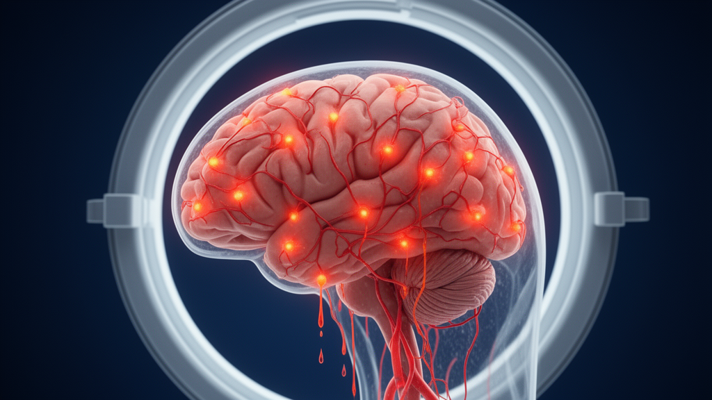
People with Parkinson’s disease show measurable signs of blood-brain barrier dysfunction on advanced MRI, according to an exploratory cross-sectional study published in Fluids and Barriers of the CNS. Using dynamic contrast-enhanced magnetic resonance imaging (DCE-MRI), researchers found that patients with Parkinson’s had higher barrier permeability and altered contrast kinetics across multiple brain regions compared with age-matched controls, along with distinctive changes at the choroid plexus, the interface where blood is filtered into cerebrospinal fluid.
The study, led by Sapir Fajerzstein and colleagues, enrolled 14 patients with Parkinson’s disease and nine age-matched controls, all scanned on a 3-Tesla MRI scanner. The team quantified three imaging measures in predefined regions spanning the cortex, subcortical structures, white matter, midbrain, and choroid plexus: the transfer constant (Ktrans), which reflects leakage of contrast agent across the barrier; the fractional plasma volume (vp); and a normalized permeability index (NPI) designed to be sensitive to slow contrast accumulation over time.
Compared with controls, participants with Parkinson’s disease demonstrated higher Ktrans and NPI and lower vp across multiple regions, including the basal ganglia, thalamus, hippocampus, midbrain, cerebral cortex, and white matter. In plain terms, the contrast agent appeared to cross the blood-brain barrier more readily and accumulate more extensively in the patients’ brain tissue, while the proportion of blood plasma within the measured tissue was reduced.
Voxel-wise analyses, which examine the brain at the level of individual volume elements rather than predefined regions, supported the regional findings. These analyses showed increased permeability predominantly in the frontal cortex and widespread reductions in fractional plasma volume throughout the imaged brain.
The choroid plexus emerged as a particular focus of the study. In this structure, Parkinson’s disease was associated with higher bilateral Ktrans, higher NPI on the left side, and lower bilateral vp. The authors interpret these altered choroid plexus contrast kinetics as suggesting involvement of the blood-cerebrospinal fluid interface, extending the observed abnormalities beyond the blood-brain barrier itself.
Within the Parkinson’s group, the researchers explored whether imaging measures related to clinical characteristics. Longer disease duration was associated with higher NPI, and higher levodopa equivalent daily dose (LEDD) was associated with lower vp. In an exploratory analysis of seven patients with unilateral symptom onset, NPI was higher in the brain hemisphere contralateral to the clinically affected body side, an intriguing hint that barrier changes may track the lateralized nature of the disease in some patients.
Not every clinical association held up. Montreal Cognitive Assessment (MoCA) scores showed no association with any imaging parameter, and disease severity showed no association independent of LEDD. The authors stress that these within-PD analyses were exploratory and require validation in larger longitudinal cohorts.
The findings come against a background of uncertainty. As the authors note, neurovascular dysfunction may contribute to Parkinson’s disease, but the extent and clinical relevance of blood-brain barrier alterations have remained unclear. DCE-MRI offers a way to probe these questions directly in living patients, quantifying barrier permeability and plasma volume without invasive procedures.
The study is published as an open-access article under a shared early-access model, meaning the peer-reviewed, accepted version has been released with a permanent DOI ahead of final copyediting. The authors describe the work as exploratory, and the small sample size of 14 patients and nine controls limits the strength of the conclusions.
Nevertheless, the report adds to a growing body of work examining the vasculature and fluid interfaces of the brain in Parkinson’s disease. If confirmed in larger longitudinal studies, the imaging signatures described here, particularly the choroid plexus changes and the laterality finding, could help clarify how neurovascular and blood-cerebrospinal fluid barriers participate in the progression of Parkinson’s disease.
Subject of Research: Blood-brain barrier permeability and choroid plexus contrast kinetics measured with DCE-MRI in Parkinson's disease
Article Title: Blood-brain barrier dysfunction and altered choroid plexus contrast kinetics in Parkinson’s disease
Article References: Fajerzstein, S., Maayan Eshed, G., Mirloo, S., Rabinowich, A., Thaler, A., Mirelman, A., Shiner, T., Giladi, N., Alcalay, R. N., Orad, R. I., Artzi, M., Bregman, N., Kamintsky, L., Omer, N., Brenner-Ullman, A., Ben Bashat, D., & Milikovsky, D. Z. (2026). Blood-brain barrier dysfunction and altered choroid plexus contrast kinetics in Parkinson’s disease. Fluids and Barriers of the CNS. https://doi.org/10.1186/s12987-026-00890-7
Image Credits: AI Generated
DOI: 10.1186/s12987-026-00890-7
Keywords: Parkinson's disease, blood-brain barrier, choroid plexus, DCE-MRI, neurovascular dysfunction, Ktrans, fractional plasma volume, permeability, levodopa, MoCA, brain imaging, Blood-brain
News Source: Cassandra Pierce. (October 28, 2026). MRI Study Links Blood-Brain Barrier Leaks to Parkinson’s Disease. Scienmag.