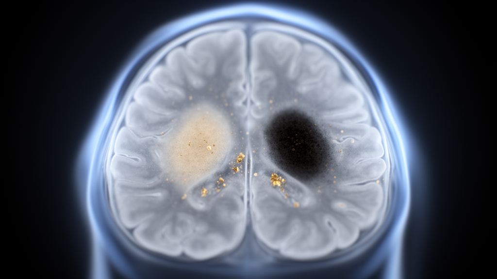
A multimodal magnetic resonance imaging study has pinpointed nigrosome-1, a small subregion of the substantia nigra pars compacta, as the main site where MRI captures the nigral degeneration associated with Parkinson’s disease. The research compared idiopathic Parkinson’s disease (iPD, n = 30), GBA-associated Parkinson’s disease (GBA-PD, n = 26) and healthy controls (HC, n = 30) using neuromelanin- and iron-sensitive MRI measures.
Parkinson’s disease involves early neuromelanin loss and iron dysregulation, and it often presents with asymmetric motor onset. The researchers quantified these features by performing magnitude-based metrics, asymmetry indices and cross-modal coupling analyses within the substantia nigra pars compacta and its nigrosome-1 subregion.
Both patient groups showed reduced neuromelanin-sensitive signal and increased iron-sensitive signal relative to healthy controls, with the strongest alterations concentrated in nigrosome-1. This pattern held across idiopathic and GBA-associated forms of the disease, indicating that the subregion carries a shared imaging signature of nigral degeneration.
Neuromelanin-sensitive nigrosome-1 markers provided the best discrimination between patients and controls. The separation was particularly strong between GBA-PD and healthy controls, reaching an area under the curve of 0.91 under leave-one-out cross-validation (AUCLOOCV = 0.91).
Nigrosome-1 neuromelanin asymmetry and, most robustly, the nigrosome-1 neuromelanin contrast ratio (CR) moderately distinguished healthy controls from the patient subgroups, with AUCLOOCV values in the range of 0.71 to 0.72. These contrast-based measures therefore performed less strongly than the overall neuromelanin-sensitive marker set but still carried patient-control information.
Asymmetry index (AI) metrics did not differentiate idiopathic Parkinson’s disease from GBA-associated Parkinson’s disease. Despite the frequent clinical observation of asymmetric motor onset in Parkinson’s disease, the MRI-derived asymmetry measures in this cohort could not separate the two patient groups from each other.
The study also examined cross-modal coupling between neuromelanin and iron signals. The neuromelanin-iron coupling pattern observed in healthy controls was inverted in Parkinson’s disease, suggesting that the normal relationship between these two tissue properties breaks down in the affected nigral tissue.
Exploratory analyses suggested a GBA1 severity-related variation in nigrosome-1 neuromelanin-iron coupling, hinting that the strength of this coupling may track genetic disease burden in GBA-associated cases. The authors note that these findings identify nigrosome-1 as the main subregion where multimodal MRI captures Parkinson’s disease-related nigral degeneration. The work was published open access in npj Parkinson’s Disease on 28 October 2026, and the reported version is a shared early release that remains subject to further editorial edits before the final Version of Record.
Subject of Research: Neuromelanin- and iron-sensitive MRI of the substantia nigra and nigrosome-1 in idiopathic and GBA-associated Parkinson's disease
Article Title: Nigrosome-1 neuromelanin and iron coupling in idiopathic and GBA-associated Parkinson disease
Article References: Martínez, M., Ariz, M., Alvarez, I., Santoro, T. M., Castellanos, G., the Catalonian Neuroimaging Parkinson’s disease Consortium, Pascual-Sedano, B., Caballol, N., Avila, A., Aguilar, M., Buongiorno, M., Tartari, J. P., Puente, V., Ezquerra, M., Garrido, A., Marti, M. J., Valldeoriola, F., Tolosa, E., Pont, C., … Pastor, P. (2026). Nigrosome-1 neuromelanin and iron coupling in idiopathic and GBA-associated Parkinson disease. npj Parkinson's Disease. https://doi.org/10.1038/s41531-026-01599-y
Image Credits: AI Generated
DOI: 10.1038/s41531-026-01599-y
Keywords: Parkinson's disease, nigrosome-1, substantia nigra, neuromelanin, iron, MRI, GBA, asymmetry index, contrast ratio, cross-modal coupling, biomarkers, neuroimaging
News Source: Diana Fleming. (October 28, 2026). MRI reveals nigrosome-1 neuromelanin and iron changes in Parkinson disease. Scienmag.