In a groundbreaking advancement poised to transform prognostic strategies for hepatocellular carcinoma (HCC), researchers have unveiled a sophisticated predictive model harnessing the power of multiparametric magnetic resonance imaging (MRI) combined with cutting-edge radiomics and clinical data. This innovative study focuses on patients who have undergone postoperative adjuvant transarterial chemoembolization (PA-TACE), a commonly employed intervention aimed at mitigating tumor recurrence following surgical resection. The study’s findings indicate a significant leap toward personalized cancer care by accurately anticipating recurrence-free survival (RFS) in this challenging patient population.
Hepatocellular carcinoma remains a formidable clinical challenge globally due to its high recurrence rates even after curative treatments such as surgical resection. Although transarterial chemoembolization serves as an adjuvant therapy to improve outcomes postoperatively, recurrence prediction remains imprecise, complicating follow-up and treatment planning. Against this backdrop, the development of a reliable predictive tool using noninvasive imaging biomarkers offers a potential paradigm shift in managing postoperative HCC.
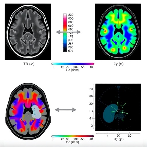
At the heart of this research lies the application of radiomics, an emerging field that extracts a vast array of quantitative features from medical images, revealing subtle information beyond what is visually perceptible. This study capitalized on multiparametric MRI, acquiring multiple sequences that capture diverse biological characteristics of tumor tissue. By integrating these complex imaging features using advanced computational strategies, the investigators sought to distill an imaging signature predictive of tumor recurrence.
The research was conducted retrospectively across two medical institutions involving 149 patients with confirmed HCC treated by PA-TACE. This cohort was methodically divided into training, internal validation, and external validation groups, ensuring the robustness and generalizability of the results. The use of multiple centers not only adds credibility but also reflects the model’s potential applicability across diverse clinical settings.
Radiomics features were meticulously extracted from three distinct MRI sequences. Each sequence provides unique tissue contrasts, capturing information on tumor heterogeneity and microenvironment alterations linked with aggressive disease behavior. The high-dimensional data extracted required sophisticated selection methods to prevent overfitting and identify the most prognostically relevant features, a task accomplished using the Least Absolute Shrinkage and Selection Operator Cox regression (LASSO-COX).
The LASSO-COX technique, known for its ability to handle multicollinearity and high-dimensional predictors, narrowed down an expansive feature set to 15 optimal radiomic variables. These features, when combined into a singular Rad-score, represented a quantitative imaging biomarker predictive of recurrence risk. The median Rad-score established a threshold distinguishing patients with divergent prognoses following PA-TACE treatment.
Beyond imaging, clinical parameters played a pivotal role in refining the prediction model. Among the numerous clinical variables analyzed, the neutrophil-to-lymphocyte ratio (NLR) and tumor size emerged as independent predictors significantly associated with recurrence-free survival. The NLR, a marker of systemic inflammation, has gained attention for its prognostic relevance across malignancies, reflecting the complex interplay between tumor biology and host immune response.
Tumor size, a long-recognized prognostic factor, reflects tumor burden and likelihood of occult metastases or vascular invasion, both of which portend a higher recurrence probability. Integrating these clinical parameters with the Rad-score yielded a composite nomogram, a user-friendly tool that transforms complex predictive analytics into individualized risk assessments.
Performance metrics underscored the model’s predictive strength. The concordance indices (C-index) measuring agreement between predicted and actual outcomes consistently exceeded 0.82 across training and validation cohorts, highlighting the model’s discriminative power. Receiver operating characteristic (ROC) curves elucidated the time-dependent accuracy of the model, substantiating its potential utility in clinical decision-making.
Calibration curves further demonstrated excellent concordance between predicted probabilities and observed recurrence rates, an essential attribute for any predictive model aspiring to clinical integration. This alignment suggests that clinicians can rely on the nomogram’s outputs to enhance postoperative surveillance strategies and potentially guide adjuvant treatment decisions tailored to individual patient risk profiles.
The implications of this study extend beyond simple prognostication. By leveraging multiparametric MRI—a noninvasive, widely available imaging modality—alongside computational radiomics, this model offers a precision medicine approach that can dynamically capture tumor biology. Such insight can facilitate early intervention at recurrence and potentially improve long-term survival outcomes for HCC patients.
Furthermore, the study exemplifies how interdisciplinary collaboration between radiology, oncology, computational science, and clinical epidemiology converges to address complex oncological challenges. It underscores the importance of integrating diverse data streams to forge predictive tools that transcend traditional staging systems, which often fail to encapsulate tumor heterogeneity and host factors adequately.
While the study’s retrospective design introduces inherent limitations, the inclusion of an external validation cohort strengthens confidence in the model’s applicability across institutions. Future prospective studies and clinical trials will be imperative to confirm these findings and evaluate how this radiomics-based nomogram can be incorporated into routine clinical workflows.
In the era of artificial intelligence and big data, the successful application of radiomics for personalized oncologic prognosis heralds a new chapter for imaging biomarkers. This research not only underscores the transformative potential of multiparametric MRI but also paves the way for radiomics-driven tools to become integral components of cancer management paradigms worldwide.
Ultimately, these advancements are poised to optimize postoperative management of hepatocellular carcinoma by enabling clinicians to identify high-risk patients who might benefit from intensified surveillance or adjunctive therapies. Such precision-guided care promises to improve patient quality of life and eventual survival through timely and targeted interventions.
As the oncology community continues to embrace innovative methodologies, studies like this provide invaluable blueprints demonstrating how quantitative imaging data can revolutionize traditional clinical prognostication. This holistic approach represents a critical step forward in realizing personalized medicine for liver cancer patients.
The confluence of multiparametric imaging and radiomics with robust clinical variables transforms the landscape of postoperative HCC care. With ongoing refinement and validation, such models hold immense promise for broader oncology applications, ultimately leading to more informed, data-driven therapeutic decisions.
Researchers and clinicians eagerly await further developments and validation studies to unlock the full potential of radiomics-based nomograms in hepatocellular carcinoma and beyond. This landmark study exemplifies the burgeoning synergy between technology and medicine aiming to improve outcomes in one of the world’s most challenging malignancies.
Subject of Research: Prediction of hepatocellular carcinoma recurrence after postoperative adjuvant transarterial chemoembolization using multiparametric MRI-based radiomics combined with clinical data.
Article Title: Multiparametric MRI-based radiomics and clinical nomogram predicts the recurrence of hepatocellular carcinoma after postoperative adjuvant transarterial chemoembolization.
Article References:
Guo, X., Song, J., Zhu, L. et al. Multiparametric MRI-based radiomics and clinical nomogram predicts the recurrence of hepatocellular carcinoma after postoperative adjuvant transarterial chemoembolization. BMC Cancer 25, 683 (2025). https://doi.org/10.1186/s12885-025-14079-y
Image Credits: Scienmag.com
DOI: https://doi.org/10.1186/s12885-025-14079-y
Tags: advanced imaging in liver cancerclinical data integration in radiomicshepatocellular carcinoma recurrence predictionimaging biomarkers in oncologyinnovative approaches to cancer managementMRI radiomics for liver cancermultiparametric MRI techniquesnoninvasive cancer recurrence assessmentpersonalized cancer care strategiespostoperative adjuvant transarterial chemoembolizationpredictive modeling in hepatocellular carcinomatumor recurrence risk factors

