Friedreich ataxia represents the most prevalent inherited ataxia with early onset, primarily manifesting during childhood or adolescence. This neurodegenerative disorder is characterized by progressive damage in specific regions of the central nervous system (CNS), with distinct anatomical changes observable through advanced imaging techniques. Gross anatomical evaluation reveals pronounced atrophy in the spinal cord and medulla oblongata, key components of the brainstem, which regulate vital bodily functions and motor coordination. Less overt yet clinically significant are the degenerative changes seen in the dentate nucleus and the superior cerebellar peduncle (SCP), structures integral to cerebellar output and motor control.
Intriguingly, early to moderate stages of Friedreich ataxia often exhibit minimal changes in brain and cerebellar volume when observed via routine clinical examination, yet sophisticated volumetric and diffusion imaging modalities reveal abnormalities throughout the ascending somatosensory pathways. These pathways, including the spinocerebellar and cerebello-thalamo-cortical tracts, facilitate the transmission of sensory input necessary for motor coordination. Although cerebellar cortical atrophy is largely absent in the initial disease phases, longitudinal studies suggest that neurodegeneration becomes evident as the disorder progresses, underscoring the chronic and progressive nature of Friedreich ataxia.
Magnetic resonance imaging (MRI) has become central to evaluating Friedreich ataxia, focusing on quantifying volumetric changes in the spinal cord, cerebellum—especially the dentate nucleus—and SCP. There is growing consensus within the scientific community that the disease entails both impaired neurodevelopmental growth and ongoing neurodegeneration, with most pronounced growth deficits observed within the spinocerebellar tract. This dual impact complicates the interpretation of imaging findings but also provides targets for monitoring disease trajectory and therapeutic response.
Initial MRI investigations from the 1990s relied primarily on manual measurements and atrophy rating scales focused on the spinal cord and medulla oblongata. These methods laid the groundwork for the transition into volumetric brain studies in the 2000s. It was not until the following decade that effect sizes for regional brain volume changes were systematically reported. Recent cross-sectional studies have quantified atrophy relative to healthy controls, consistently demonstrating large effect sizes in spinal cord and brain structures, particularly the medulla, SCP, inferior cerebellar peduncle (ICP), and cervical spinal cord cross-sectional area (CSA). Remarkably, these volumetric alterations are already present in ambulatory patients with early-stage disease, indicating an early and robust impact of Friedreich ataxia on CNS structures.
Longitudinal MRI studies, although fewer, provide significant insight into disease progression. Among adult patients, volumes of the total cerebellum and brainstem emerge as sensitive markers for monitoring changes over time, followed closely by SCP volume and cervical spinal cord CSA, which also show measurable longitudinal degeneration. However, some regions with large cross-sectional effect sizes, such as the medulla, have not demonstrated statistically significant longitudinal atrophy, likely due to the small proportion of affected ascending pathways in this region and technical limitations related to physiological motion during imaging.
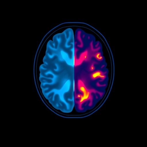
Beyond simple volumetric measurements, advanced MRI techniques such as diffusion MRI (dMRI), magnetic resonance spectroscopy (MRS), and quantitative susceptibility mapping (QSM) have expanded our understanding of the microstructural and biochemical brain changes in Friedreich ataxia. High cross-sectional effect sizes have been reported for dMRI parameters, particularly fractional anisotropy and radial diffusivity, in the SCP and cervical spinal cord. These metrics reflect white matter integrity and are sensitive markers of microstructural disruption. QSM has provided valuable in vivo insights by mapping changes in magnetic susceptibility within the dentate nucleus, a proxy for iron accumulation, myelin loss, and inflammation. Similarly, MRS data reveal alterations in neurochemical ratios in the cervical spinal cord, further supporting the multifaceted impact of Friedreich ataxia on neural tissue.
Importantly, these advanced imaging biomarkers exhibit correlations with clinical outcome assessments (COAs), reinforcing their clinical relevance. Measures such as SCP volume, brainstem volume, and cervical spinal cord CSA correlate with disease severity scales like the Friedreich Ataxia Rating Scale (FARS) and the Scale for the Assessment and Rating of Ataxia (SARA). Diffusion metrics showing reduced fractional anisotropy and increased radial diffusivity align with poorer clinical function, while QSM parameters of iron-related susceptibility in the dentate nucleus correspond to higher disability scores. Such correlations suggest these imaging metrics can serve as surrogate markers for clinical decline and potential endpoints in therapeutic trials.
Despite these advances, substantial gaps remain in the current knowledge base. Most studies have involved relatively small cohorts and have been conducted at single sites, limiting the ability to parse variability related to patient age, stage of disease, or symptom onset timing, such as pediatric versus adult cases. Large, multimodal, longitudinal studies remain sparse, though ongoing efforts through consortia like ENIGMA-Ataxia and TRACK-FA are expected to provide valuable data soon. Additionally, how imaging biomarkers perform in terms of sensitivity and specificity across different clinical phases of Friedreich ataxia is not definitively established. This is particularly pertinent given that disease progression rates and imaging changes may follow nonlinear trajectories.
A crucial but underexplored area relates to the early, pre-symptomatic development of CNS abnormalities in Friedreich ataxia. Since newborn genetic screening is not yet routine for this recessive disorder, data on very young or asymptomatic individuals are limited. Nevertheless, preliminary cross-sectional data from the TRACK-FA study indicate that CNS differences between affected individuals and controls arise in early childhood, emphasizing the potential of imaging techniques to identify early disease markers. Future incorporation of newborn screening, combined with presymptomatic imaging, may pave the way for preventative clinical trials aiming to delay or mitigate disease onset.
Recommended neuroimaging endpoints for clinical trials in Friedreich ataxia focus on metrics with high sensitivity and clarity. SCP volume, medulla oblongata volume, and cervical spinal cord CSA exhibit large cross-sectional effect sizes and are indicative of impaired developmental growth, making them valuable early disease markers. For monitoring progression in adults, total cerebellum volume and brainstem volume are preferred for their superior longitudinal sensitivity, while SCP volume and upper cervical CSA remain important though with somewhat reduced longitudinal effect sizes. Furthermore, diffusion MRI measures within the SCP and quantitative susceptibility mapping in the dentate nucleus show promise as markers for tracking disease progression and for early detection of therapeutic response.
Clinical trials have begun leveraging these biomarkers to evaluate treatment efficacy. For instance, a small randomized controlled trial investigating leriglitazone, a brain-penetrant PPARγ agonist, revealed that patients treated with the drug exhibited a significantly attenuated increase in dentate nucleus susceptibility over 48 weeks compared to placebo. This finding suggests that leriglitazone may slow pathological iron accumulation or related neurodegenerative processes in affected brain regions, highlighting the potential of advanced MRI biomarkers to detect subtle therapeutic effects.
While structural MRI-based monitoring has ushered in new opportunities for understanding Friedreich ataxia, challenges persist concerning standardization. Variability in acquisition protocols, imaging hardware, and data processing pipelines between research groups complicates direct comparison of results across studies. Establishing universal standards and harmonized methodologies will be essential to maximize the utility of imaging biomarkers and to facilitate multi-center clinical trials. The large-scale, longitudinal TRACK-FA initiative is poised to provide critical data to address these concerns and refine imaging endpoints with stronger clinical correlations.
Emerging evidence from quantitative neuroimaging underscores the intricate interplay between impaired neurodevelopment and progressive neurodegeneration in Friedreich ataxia. This dual process manifests as reduced growth of specific neural pathways early in life, along with sustained atrophic changes throughout disease progression. Key target regions such as the spinal cord, brainstem, SCP, and dentate nucleus represent structural and biochemical hubs where pathological processes converge. Improved understanding of these mechanisms through advanced imaging not only enhances diagnostic accuracy but also informs therapeutic targeting and timing.
The impact of Friedreich ataxia spans sensory, motor, and autonomic systems, reflecting widespread CNS involvement beyond classical cerebellar atrophy. Volumetric and microstructural alterations in ascending somatosensory and motor pathways highlight the extensive degenerative cascade. This complexity necessitates multiparametric imaging frameworks that combine volumetry, diffusion metrics, spectroscopic data, and susceptibility analyses to provide a comprehensive picture. Integrating such multiparametric data in clinical trials offers the potential to detect disease-modifying effects with maximal sensitivity and specificity.
Looking forward, the integration of neuroimaging biomarkers with genetic, biochemical, and clinical data will pave the way for personalized medicine approaches in Friedreich ataxia. Early detection of subclinical changes could enable stratification of patients for tailored interventions, while imaging endpoints might serve as surrogate markers for monitoring therapeutic efficacy in modify neuroprotective or disease-modifying trials. The ongoing advancement of high-resolution imaging techniques and computational analytics promises to transform Friedreich ataxia management and clinical trial design fundamentally.
In conclusion, MRI-based biomarkers are rapidly evolving as indispensable tools in the understanding and treatment of Friedreich ataxia. Despite substantial progress, challenges such as cohort heterogeneity, limited pediatric and presymptomatic data, and standardization barriers remain. Addressing these limitations through large-scale longitudinal studies and harmonized protocols is critical. Continued validation of volumetric, diffusion, spectroscopic, and susceptibility imaging outcomes as clinically meaningful surrogates will support their adoption as primary endpoints in clinical trials. Such advances are essential to accelerate therapeutic development and ultimately improve outcomes for individuals affected by this debilitating genetic ataxia.
Subject of Research: Neurodegenerative changes and MRI biomarkers in Friedreich ataxia
Article Title: MRI end-points for clinical trials in ataxias: recommendations from the Ataxia Global Initiative MRI Biomarkers Working Group
Article References:
Öz, G., Cocozza, S., Rezende, T.J.R. et al. MRI end-points for clinical trials in ataxias: recommendations from the Ataxia Global Initiative MRI Biomarkers Working Group. Nat Rev Neurol (2026). https://doi.org/10.1038/s41582-026-01218-7
Tags: advanced diffusion imaging for ataxiacerebellar peduncle degenerationcerebello-thalamo-cortical pathway imagingdentate nucleus abnormalitiesearly-stage Friedreich ataxia biomarkersFriedreich ataxia neurodegenerationlongitudinal MRI studies in neurodegenerative disordersMRI endpoints for ataxia clinical trialsMRI volumetric analysis in ataxianeuroimaging in inherited ataxspinal cord atrophy imagingspinocerebellar tract assessment


