


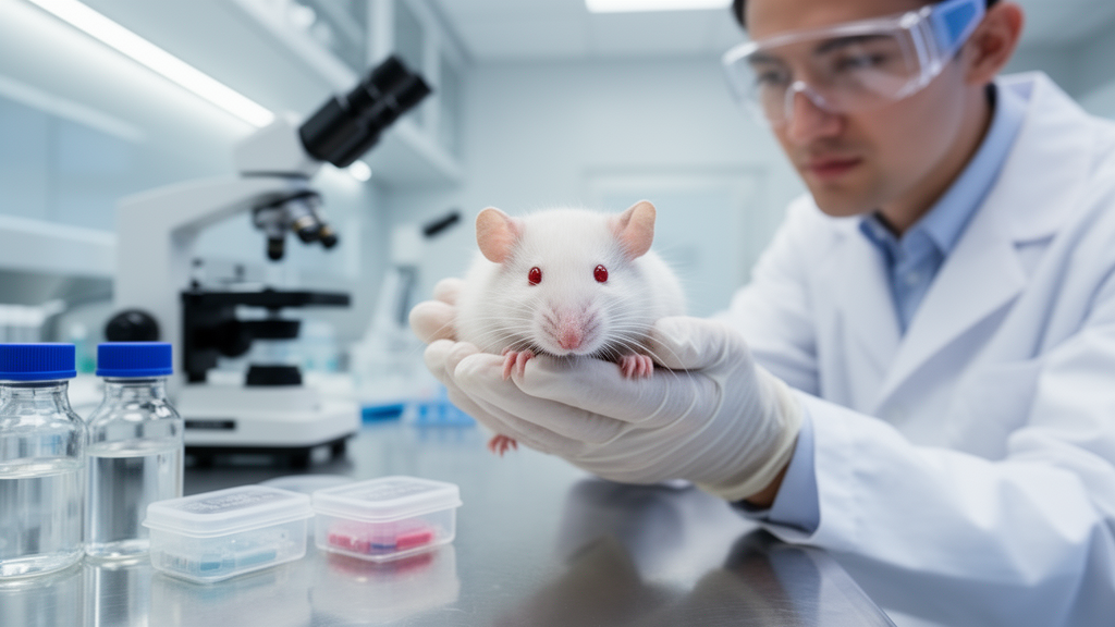
Chandipura virus remains one of India’s most troubling pediatric pathogens, yet almost everything scientists know about how it kills has come from a single type of experimental animal. A comprehensive review published in Discover Viruses on 27 February 2026 examines the laboratory models used to study the virus and concludes that mice, particularly neonatal and juvenile animals, have carried nearly the entire burden of Chandipura virus research, while studies in other species remain sparse and largely limited to antibody surveys. The review, authored by Thuruthiyil Ramachandran Divya, Srinivas Vilasagaram, Mukesh Kumar Gupta and Ravindran Kumar Pramod, analyses how these models have shaped understanding of viral tropism, immune responses and disease mechanisms, and where they fall short of reproducing natural infection.
Chandipura virus is a neurotropic arbovirus of the family Rhabdoviridae, genus Vesiculovirus, first isolated in 1965 during an outbreak in Chandipura village in Maharashtra, India, from adults presenting with fever and joint pain. The virus subsequently became associated with recurring outbreaks of acute encephalitis disease in India, primarily affecting children and producing significant mortality. Across seven outbreaks reported between 2003 and 2024, the case fatality rate ranged from 28 percent to 79 percent. The disease primarily affects children below 15 years of age and is characterised by febrile onset that can progress to neurological symptoms including convulsions, coma and, in some cases, death. Notable outbreaks have included a large encephalitis event in Andhra Pradesh from June to September 2003 affecting 55 children aged 2.5 months to 15 years, followed by outbreaks in Gujarat in 2004, Andhra Pradesh in 2005-06, Maharashtra in 2007, Odisha in 2009 and Bihar in 2016. Most recently, Gujarat reported an outbreak involving 148 cases of acute encephalitis syndrome in children under 15, of which 51 were laboratory-confirmed as Chandipura virus.
Despite this public health burden, there are currently no specific antiviral treatments or licensed vaccines for Chandipura virus infection, a gap the review identifies as the central motivation for developing and refining experimental models. Animal models serve as surrogates for human infection, allowing researchers to track viral spread within an organism, identify target organs, and evaluate antiviral compounds and vaccine candidates for immunogenicity, protective efficacy, safety and dosage before human trials. The review argues that establishing truly representative models is crucial for studying pathogenesis and for preclinical testing, and it critically evaluates the strengths and limitations of rodents and other species in recapitulating different aspects of human infection.
Laboratory mice have traditionally been the primary model for studying Chandipura virus pathophysiology, and the pattern of susceptibility they display is strikingly age-dependent. Neonatal and young mice, whose blood-brain barrier is immature, are vulnerable to infection when inoculated via intravenous and intraperitoneal routes and show neurobehavioral symptoms that closely mimic human disease. Adult mice, with a fully developed barrier, show clinical symptoms only upon intracranial inoculation. Experimental infection has been established in several strains, including inbred BALB/c and C3H mice, outbred CD1 and Swiss albino stocks, and immunocompromised lines such as severe combined immunodeficiency and athymic nude mice. Neonatal and young mice can be infected by footpad, intraperitoneal, intravenous, subcutaneous and intramuscular routes, while adults require the intracranial route. The cause of these age-dependent differences is not known but may reflect protection by innate immunity in young animals or prevention of viral entry into the brain by a fully mature barrier in adults. In mice, the blood-brain barrier becomes functionally mature around embryonic day 15.5.
The foundations of murine work were laid in 1973, when Jortner and co-workers inoculated nine-day-old Swiss mice with 4.2 log10 TCID50 of Chandipura virus strain 16,535,141 and documented the virus’s neurotropic and age-dependent lethality. A 1986 study by Wilks and House, using the Rockefeller H/Plum Island strain at the Plum Island Animal Disease Center, categorised mice into day-old neonates and two adult groups. Day-old mice succumbed to infection following both intraperitoneal and intracranial inoculation, with viral titres reaching 10^12.9 TCID50 per gram in brain tissue, whereas adult mice exhibited resistance regardless of administration route. Mice can also be infected via experimentally infected sand flies: Mavale and co-workers, assessing vector competence, housed intrathoracically inoculated female flies with pairs of baby mice and observed that, with a fly feeding rate of 63 percent and within-fly viral dissemination of 93 percent, the average fly-to-mouse transmission rate across groups was 32 percent.
Studies in young mice have mapped the course of infection in detail. Following intravenous inoculation, researchers demonstrated rapid viral invasion of the central nervous system, with peak brain titres at 72 hours post-infection and death occurring by 72 to 96 hours. Seroconversion to virus-specific IgM occurred by 48 hours, peaking at 72 hours, but no virus-specific IgG antibodies were detected. Mice passively immunised with anti-Chandipura antibodies survived, suggesting a protective role for neutralising antibodies. The same work recorded significant reductions in CD4-positive, CD8-positive and CD19-positive cell populations along with antigen-specific suppression of T cell proliferation, while TNF-alpha levels spiked at 24 hours post-infection before declining. Virus-specific CD4-positive regulatory T cells have also been reported during infection, potentially influencing activated lymphocytes through unique regulatory mechanisms. Anukumar and Shahir demonstrated in 2012 that the virus activates Toll-like receptor 4 signalling pathways, leading to induction of proinflammatory cytokines, using TLR4-mutant and wild-type mice. In 2013, Anukumar and colleagues showed that footpad inoculation of 13-day-old Swiss albino mice produced viral RNA predominantly in nervous tissue at 96 hours, with transient replication in non-neural tissues declining after 24 hours, alongside hindlimb paralysis, circling movements, eye lesions and spinal cord pathology. Ghosh and co-workers confirmed selective neuronal replication in 10-day-old BALB/c mice, identified cholesterol as a critical host factor, and later reported elevated inducible nitric oxide synthase, cyclooxygenase-2, nitric oxide and reactive oxygen species in infected brains.
Murine models have also served as the testing ground for countermeasures. Venkateswarlu and Arankalle developed a recombinant vaccine using the complete G gene from a patient isolated during the 2003 Andhra Pradesh outbreak, expressed with a baculovirus system. A three-dose schedule four weeks apart achieved 90 percent seroconversion and significant protection against challenge with live virus at 100 LD50, with neutralising antibodies detectable as early as two weeks after the first dose and 60 percent T-cell proliferation against the recombinant glycoprotein. The same team showed that combining the candidate with a commercially available diphtheria, tetanus and pertussis vaccine achieved 90 to 100 percent seroconversion, high ELISA titres of 1:1200 to 1:2400, stable antibody levels for six months and protection against intracerebral challenge. A Vero cell-based candidate evaluated in four-week-old Swiss albino mice produced dose-dependent antibody titres, with the third dose eliciting the highest neutralising activity. On the therapeutic side, an siRNA targeting the P gene, delivered with cationic lipid in two 5-microgram doses at 0 and 24 hours post-infection, achieved a 70 percent survival rate, delayed mortality and reduced viral load by 4 log, with no detectable anti-Chandipura IgG in surviving mice at 7, 14 and 21 days. Minocycline, given daily at 22.5 mg/kg body weight for three days starting 8 hours post-infection, attenuated cytoplasmic calcium accumulation, reduced reactive oxygen species generation and inhibited p38 MAPK phosphorylation, extending survival by 3 to 4 days beyond untreated controls that died 3 days post-infection, though it could not prevent death. In a severe combined immunodeficiency model, favipiravir improved survival with both pre-symptomatic and post-symptomatic regimens and reduced viral loads in blood, kidney and adrenal tissue and brain.
Non-murine species tell a very different story. The 1986 Wilks and House assessment covered ponies, steers, sheep, goats, pigs, hamsters, guinea pigs and rabbits. Two of four ponies developed vesicular lesions on the tongue and mouth, with infectious virus up to 10^8.1 TCID50 per gram recovered from tongue tissue and neutralising antibodies by 7 to 14 days. One of two steers developed shallow tongue ulcers with virus recovered from lesions, and goats showed shallow ulcers in one of three animals with neutralising antibodies in all. Sheep showed no visible lesions or detectable virus, though two of three had precipitating antibodies by 29 days. Pigs showed no lesions or virus isolation, but all developed neutralising antibodies, while contact pigs did not seroconvert significantly. Guinea pigs mounted neutralising antibodies without disease; day-old hamsters died within 24 hours of intraperitoneal or intracranial inoculation while adults resisted infection yet still produced neutralising antibody; and rabbits showed no clinical signs but mounted virus-neutralising titres above 360. The review notes that outside mice, studies have been largely restricted to serosurveillance, with anti-Chandipura antibodies documented in pigs, buffaloes, cattle, goats and sheep, and limited evidence of overt susceptibility.
The review’s conclusions are measured. Mouse models have been indispensable for understanding neurotropism, neuronal damage and host-virus immune interplay, and have facilitated preclinical evaluation of favipiravir and vaccine candidates showing promising results that warrant further investigation for translation into human clinical applications. Yet their limitations are real: age-dependent susceptibility means adult models frequently require intracranial inoculation to achieve fatal infection, which may fail to represent natural transmission pathways, and the precise mechanisms of neuroinvasion and neuronal death still require clarification. The authors call for physiologically relevant models replicating natural infection routes across age groups, systematic exploration of non-murine species with optimised inoculation parameters, investigation of the cytokine storm observed in severe cases, combination therapy testing, identification of early biomarkers, and establishment of vaccine correlates of protection to guide development of effective human vaccines.
Subject of Research: Animal models used in Chandipura virus research
Article Title: Animal models for Chandipura virus (CHPV) research: a comprehensive review
Article References: Divya, T. R., Vilasagaram, S., Gupta, M. K., & Pramod, R. K. (2026). Animal models for Chandipura virus (CHPV) research: a comprehensive review. Discover Viruses, 3(1), Article 3. https://doi.org/10.1007/s44370-026-00039-y
Image Credits: AI Generated
DOI: 10.1007/s44370-026-00039-y
Keywords: Chandipura virus, animal models, laboratory mice, blood-brain barrier, neuroinvasion, vaccine candidates, favipiravir, siRNA therapy, minocycline, acute encephalitis, India, Rhabdoviridae
News Source: Kristina Jarvis. (October 30, 2026). Mouse models anchor Chandipura virus research as vaccine and drug candidates advance. Scienmag.



