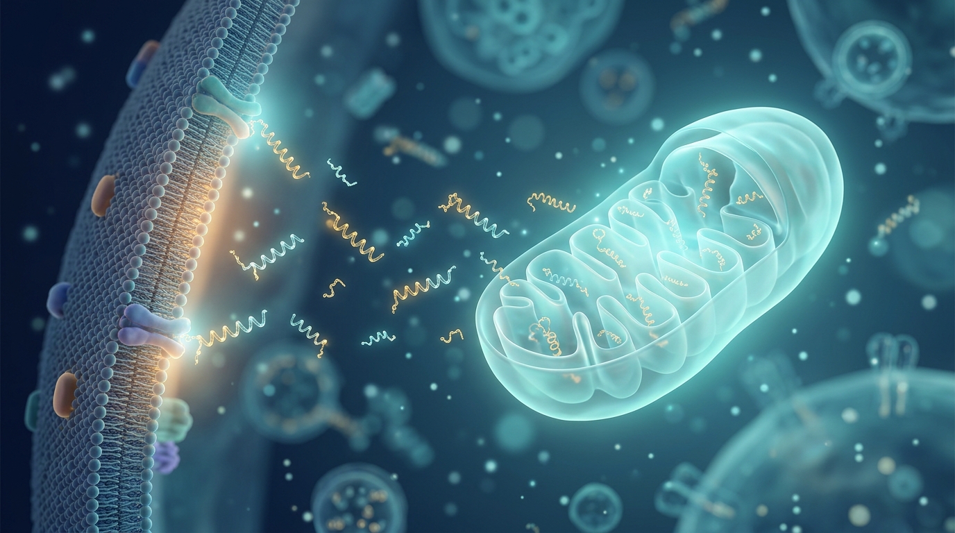
A quiet revolution in pain research is unfolding inside the cell’s smallest compartment. Mitochondria, long appreciated as the power plants that fuel neurons, also manufacture their own chemical messengers, and a growing body of preclinical evidence suggests these mitochondrial-derived peptides could become a new class of analgesic drugs. A mini review published in Frontiers in Pain Research on 30 October 2026 by Zhe Wang, Jinglei Liao, Ping Lu, Yimin Xu and Jinhong Jiang, with Zhe Wang at Xuzhou University of Technology and the other four authors at Xuzhou Medical University, synthesizes this emerging field, arguing that peptides such as humanin and MOTS-c attack chronic pain at a level most current drugs never touch: the failing mitochondrion itself.
The rationale rests on a convergent pathological signature. Whether pain arises from diabetic neuropathy, chemotherapy toxicity, traumatic nerve injury or inflammation, the same mitochondrial damage appears downstream: excessive reactive oxygen species, impaired mitochondrial biogenesis, disrupted dynamics and the activation of neuroinflammatory signaling. Existing medications, including opioids, gabapentinoids, tricyclic antidepressants and serotonin-norepinephrine reuptake inhibitors, act on a limited set of neural targets and help only a subset of patients, often with dose-limiting side effects such as dizziness, drowsiness and weight changes. Restoring mitochondrial homeostasis, the authors contend, could offer benefits across pain types that are otherwise mechanistically distinct.
Mitochondrial-derived peptides differ from conventional mitochondrial-targeted antioxidants such as MitoQ, which passively scavenge reactive oxygen species. Instead, these endogenous peptides actively reprogram cellular metabolism and stress responses. Three families have drawn particular attention: humanin, a 24-amino-acid peptide encoded by the mitochondrial 16S rRNA region; MOTS-c, a 16-amino-acid peptide encoded by the 12S rRNA region whose N-terminal sequence is highly conserved across species; and the small humanin-like peptides, or SHLPs, six members also encoded by the 16S rRNA region. All enhance mitochondrial biogenesis while suppressing oxidative stress and inflammation, positioning them as signaling molecules rather than simple radical sponges.
Humanin, the pioneering member of the family, was first characterized as a neuroprotective agent in Alzheimer’s disease models, where it suppresses amyloid-beta-induced toxicity through PI3K/AKT signaling. Its analgesic credentials come from a recent study spanning three mechanistically distinct neuropathic pain models: streptozotocin-induced diabetic neuropathy, oxaliplatin-induced chemotherapy neuropathy and sciatic nerve cuffing-induced traumatic neuropathy. Humanin at 4 milligrams per kilogram, given intraperitoneally over 15 days, significantly increased pain thresholds in all three models as measured by hot plate, cold plate and von Frey tests. In diabetic mice, chronic treatment also reduced elevated expression of pain-related markers in dorsal root ganglia, including the NMDA receptor, substance P, TRPV1 and interleukin-1beta, nodes that respectively mediate central sensitization, nociceptive neurotransmission, thermal and inflammatory transduction, and neuroinflammation.
At the systemic level, humanin administration in these neuropathic pain models increased antioxidant enzyme levels and anti-inflammatory cytokines while reducing oxidative stress markers and pro-inflammatory cytokines in serum, a profile that complements the local effects in dorsal root ganglia. Yet important questions remain open. Because humanin improves insulin sensitivity and glucose metabolism in diabetic models, improved glycemic control could indirectly reduce neuropathic pain; however, its efficacy in non-diabetic models such as oxaliplatin-induced and cuffing-induced neuropathy argues against a purely metabolic mechanism. Current mechanistic evidence also focuses almost exclusively on neurons, even though microglia and astrocytes are established drivers of neuropathic pain. Since humanin reduces interleukin-1beta, a cytokine produced predominantly by activated microglia, glial mechanisms are likely involved but have not been formally tested.
MOTS-c carries the broadest and most direct pain evidence. In a spared nerve injury model, the first direct demonstration, MOTS-c levels were significantly decreased in plasma and spinal dorsal horn, suggesting that endogenous depletion may contribute to pain pathogenesis. Intrathecal MOTS-c produced dose-dependent antinociception linked to inhibition of spinal microglial activation and reduced pro-inflammatory cytokine production. Subsequent work extended these findings to painful diabetic neuropathy, where MOTS-c delivered intrathecally, intraperitoneally or intraplantarly significantly improved mechanical allodynia and thermal hyperalgesia in streptozotocin-induced mice. Mechanistically, the peptide enhanced AMPKalpha1/2 phosphorylation and PGC-1alpha expression in the lumbar spinal cord, accompanied by restored mitochondrial biogenesis, inhibited microglial activation and decreased pro-inflammatory factor production. Efficacy has also been shown in cancer-induced bone pain through AMPK-mediated mitochondrial biogenesis, and in the formalin test of acute inflammatory pain through central and peripheral anti-inflammatory actions.
The breadth of MOTS-c’s activity across neuropathic, diabetic, inflammatory and cancer-related pain likely reflects the central role of the AMPK-PGC-1alpha signaling axis in cellular stress responses. Under resting conditions MOTS-c resides mainly in mitochondria, but under metabolic stress it translocates to the nucleus in an AMPK-dependent manner, regulating nuclear gene expression to maintain cellular homeostasis. A translational milestone already exists: CB4211, a MOTS-c analog, completed a Phase 1a/1b clinical trial for nonalcoholic steatohepatitis and obesity under registration NCT03998514. The analog was well tolerated, and after four weeks of treatment liver enzymes and blood glucose levels were significantly reduced, with measurable reductions in liver fat. That trial was not designed for pain endpoints, but it established that MOTS-c-based therapeutics can be safely administered to humans, laying a foundation for future pain-focused studies.
The SHLP family remains a blank space in pain research, but its biology hints at relevance. SHLP2, the best-characterized member, has repeatedly shown anti-inflammatory and anti-apoptotic properties in non-pain models. In an oxidative stress-induced osteoblast precursor injury model, SHLP2 at 10 micromolar significantly reduced reactive oxygen species production, downregulated pro-apoptotic markers such as p53 and BAX and elevated anti-apoptotic BCL-2. In a cellular model of age-related macular degeneration, it protected retinal pigment epithelial cells from amyloid-beta toxicity and upregulated mitochondrial respiratory chain complex I expression by up to 350 percent. In an allergic asthma model, it attenuated airway inflammation by improving mitochondrial function and inhibiting ETV5/GSDMD-mediated pyroptosis. Because neuroinflammation and apoptosis are important mechanisms maintaining neuropathic pain, these properties constitute a plausible but as-yet-unverified indirect link, one the authors say warrants validation in pain models.
The review also highlights a striking biomarker angle. Endogenous MDP levels decline in pain states, and the authors propose a vicious cycle: pain-related mitochondrial dysfunction reduces endogenous peptide production, which weakens mitochondrial protection, amplifies oxidative stress and neuroinflammation, and reinforces nociceptive sensitization. Patterns of MDP decline have been systematically documented in other diseases. Transcript levels of humanin and MOTS-c in blood and plasma are significantly lower in Alzheimer's disease patients than in controls, and in separate clinical studies plasma MOTS-c was decreased in Alzheimer's disease patients while humanin was reduced in Alzheimer's disease but unchanged in type 2 diabetes, suggesting disease-specific patterns. Circulating humanin is significantly reduced in coronary artery disease and myocardial infarction, where low humanin is an independent risk factor for infarction, and MOTS-c is similarly reduced in stable coronary artery disease and can serve as an independent predictor of coronary lesions. In metabolic disease, circulating MOTS-c is significantly reduced in obese male children and adolescents and correlates with insulin resistance, and serum humanin is significantly lower in women with gestational diabetes mellitus than in controls.
No MDP-based therapeutic has yet been clinically evaluated with pain as a primary endpoint, so all analgesic evidence remains preclinical. The authors position MDPs against neighboring mitochondrial strategies. A 2026 study in Nature by Ji Ru-Rong's group at Duke University revealed that satellite glial cells in the dorsal root ganglia transfer mitochondria to sensory neurons via MYO10-driven tunneling nanotubes, a process impaired in diabetic and chemotherapy-induced neuropathic pain models, where the nanogap between satellite glial cells and neurons expands from approximately 0.1 micrometers to 1 to 4 micrometers, interrupting mitochondrial supply. Transplanting healthy donor satellite glial cells or their purified mitochondria into the dorsal root ganglia of diabetic mice significantly improved hyperalgesia and promoted intraepidermal nerve fiber regeneration. Meanwhile, a Phase 2 trial, the Mito-T2D_PPN study registered as ACTRN12624000623594, is evaluating a ubiquinone-based combination of coenzyme Q10, magnesium, alpha-lipoic acid and thiamine for type 2 diabetes-related painful peripheral neuropathy, offering a proof-of-concept opportunity for mitochondria-targeted pain therapies. The authors argue MDPs are mechanistically more targeted than exogenous supplements because they directly activate the AMPK-PGC-1alpha axis rather than passively supplying substrates. Future priorities include incorporating validated pain endpoints into clinical trials, measuring circulating MDP levels as potential biomarkers, distinguishing direct analgesic effects from indirect benefits of improved metabolic control, and exploring combination strategies with existing analgesics. Two questions will determine whether these endogenous peptides graduate from promising preclinical targets to testable clinical candidates: whether the decline in MDP levels is a driving factor or merely an accompanying phenomenon of pain, and whether their analgesic effect reflects direct restoration of mitochondrial function or indirect action through downstream anti-inflammatory and anti-apoptotic pathways.
Subject of Research: Mitochondrial-derived peptides as emerging preclinical therapeutic targets for chronic pain
Article Title: Mitochondrial-derived peptides: emerging therapeutic targets at the intersection of mitochondrial dysfunction and pain
Article References: Wang, Z., Liao, J., Lu, P., Xu, Y., & Jiang, J. (2026). Mitochondrial-derived peptides: emerging therapeutic targets at the intersection of mitochondrial dysfunction and pain. Frontiers in Pain Research, 7, Article 2006909. https://doi.org/10.3389/fpain.2026.2006909
Image Credits: AI Generated
DOI: 10.3389/fpain.2026.2006909
Keywords: mitochondrial-derived peptides, humanin, MOTS-c, SHLP2, neuropathic pain, chronic pain, mitochondrial biogenesis, AMPK-PGC-1alpha, neuroinflammation, oxidative stress, CB4211, dorsal root ganglia
News Source: Drew Townsend. (November 4, 2026). Mitochondria-made peptides emerge as preclinical analgesic candidates. Scienmag.